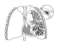
Photo from wikipedia
Objective Thermal ablation is a minimally invasive procedure for the treatment of pulmonary malignancy, but the intraoperative measure of complete ablation of the tumor is mainly based on the subjective… Click to show full abstract
Objective Thermal ablation is a minimally invasive procedure for the treatment of pulmonary malignancy, but the intraoperative measure of complete ablation of the tumor is mainly based on the subjective judgment of clinicians without quantitative criteria. This study aimed to develop and validate an intraoperative computed tomography (CT)-based radiomic nomogram to predict complete ablation of pulmonary malignancy. Methods This study enrolled 104 individual lesions from 92 patients with primary or metastatic pulmonary malignancies, which were randomly divided into training cohort (n=74) and verification cohort (n=30). Radiomics features were extracted from the original CT images when the study clinicians determined the completion of the ablation surgery. Minimum redundancy maximum relevance (mRMR) and least absolute shrinkage and selection operator (LASSO) were adopted for the dimensionality reduction of high-dimensional data and feature selection. The prediction model was developed based on the radiomics signature combined with the independent clinical predictors by multiple logistic regression analysis. The area under the curve (AUC), accuracy, sensitivity, and specificity were calculated. Receiver operating characteristic (ROC) curves and calibration curves were used to evaluate the predictive performance of the model. Decision curve analysis (DCA) was applied to estimate the clinical usefulness and net benefit of the nomogram for decision making. Results Thirteen CT features were selected to construct radiomics prediction model, which exhibits good predictive performance for determination of complete ablation of pulmonary malignancy. The AUCs of a CT-based radiomics nomogram that integrated the radiomics signature and the clinical predictors were 0.88 (95% CI 0.80-0.96) in the training cohort and 0.87 (95% CI: 0.71–1.00) in the validation cohort, respectively. The radiomics nomogram was well calibrated in both the training and validation cohorts, and it was highly consistent with complete tumor ablation. DCA indicated that the nomogram was clinically useful. Conclusion A CT-based radiomics nomogram has good predictive value for determination of complete ablation of pulmonary malignancy intraoperatively, which can assist in decision-making.
Journal Title: Frontiers in Oncology
Year Published: 2022
Link to full text (if available)
Share on Social Media: Sign Up to like & get
recommendations!