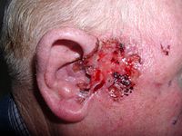
Photo from wikipedia
A woman in her 60s presented with a 3-month history of a progressive, asymptomatic cutaneous eruption on her chest and back. She had a complex history of breast cancer on… Click to show full abstract
A woman in her 60s presented with a 3-month history of a progressive, asymptomatic cutaneous eruption on her chest and back. She had a complex history of breast cancer on the left side that was first diagnosed in 1991 and was treated with lumpectomy, radiotherapy, cyclophosphamide, methotrexate, and fluorouracil. In 2001, she experienced local disease recurrence and underwent complete mastectomy with axillary lymph node dissection on the left side and received adjuvant treatment with tamoxifen citrate and anastrozole. In 2014, she developed axillary lymphadenopathy on the right side and was found to have metastatic disease. She subsequently underwent axillary lymph node dissection on the right side, followed by treatment with doxorubicin hydrochloride and cyclophosphamide. Her most recent mammogram and positron-emission tomography– computed tomography (PET/CT) showed no evidence of active disease. At the time of her evaluation, the patient’s physical examination was remarkable for discrete erysipeloid plaques on her chest, back, and right shoulder (Figure 1A). The lesions were warm and indurated but nontender to palpation. Serum chemistry profile, complete blood cell counts, and hepatic and renal function testing had normal results. Lesional skin biopsies were performed (Figure 1B). Clinical examination A Histopathologic examination B
Journal Title: JAMA oncology
Year Published: 2017
Link to full text (if available)
Share on Social Media: Sign Up to like & get
recommendations!