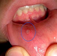
Photo from wikipedia
A woman in her 80s with a history of recurrent oral cancer presented with a new, biopsyconfirmed, squamous cell carcinoma (SCC). Her oncologic history began 16 years ago with diagnosis… Click to show full abstract
A woman in her 80s with a history of recurrent oral cancer presented with a new, biopsyconfirmed, squamous cell carcinoma (SCC). Her oncologic history began 16 years ago with diagnosis and local resection of a lesion on the palate. Cancer recurrence at the left buccal mucosa was identified and excised 10 years later. She then was diagnosed with 3 recurrences of SCC in rapid succession. Each malignant neoplasm was locally resected and the patient was also treated with adjuvant radiotherapy for the last episode. She remained asymptomatic, and a routine surveillance clinical examination uncovered a sixth lesion at the margin of a palatal graft. The patient’s medical history was also noted for a total thyroidectomy in the 1950s, complicated by recurrent laryngeal nerve injury. She had a history of tobacco use and stopped smoking cigarettes in the 1970s. Physical examination and laryngoscopy findings confirmed a painless lesion on the left side of the palate and immobility of the left vocal cord. No other mucosal lesion was identified. Positron emission tomography (PET) was performed to assess extent of disease and to plan treatment. The PET scan results showed focal fludeoxyglucose F 18 localization within the known oral cavity SCC (Figure 1A) and in a small, level IIA lymph node, located lateral to the upper right internal jugular vein. There was unexpected, prominent, metabolic activity in the medial and asymmetrically enlarged left vocal cord (Figure 1B).
Journal Title: JAMA oncology
Year Published: 2018
Link to full text (if available)
Share on Social Media: Sign Up to like & get
recommendations!