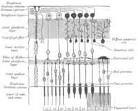
Photo from wikipedia
Importance Faster structural changes may be associated with worse vision-related quality of life in patients with glaucoma. Objectives To evaluate the association between the rate of ganglion cell complex thinning… Click to show full abstract
Importance Faster structural changes may be associated with worse vision-related quality of life in patients with glaucoma. Objectives To evaluate the association between the rate of ganglion cell complex thinning and the Vision Function Questionnaire in glaucoma. Design, Setting, and Participants This retrospective analysis of a longitudinal cohort was designed in October 2021. Patients were enrolled from the Diagnostic Innovations in Glaucoma Study and the African Descent and Glaucoma Evaluation Study. Two hundred thirty-six eyes of 118 patients with diagnosed or suspected glaucoma were followed up with imaging for a mean of 4.1 years from September 2014 to March 2020. Main Outcomes and Measures The Vision Function Questionnaire was evaluated using the 25-item National Eye Institute Visual Function at the last follow-up visit. Ganglion cell complex thickness was derived from macular optical coherence tomography scans and averaged within 3 circular areas (3.4°, 5.6°, and 6.8° from the fovea) and superior and inferior hemiregions. Linear mixed-effects models were used to investigate the association between the rate of ganglion cell complex thinning and Rasch-calibrated Vision Function Questionnaire score. Results The mean (SD) age was 73.2 (8.7) years, 65 participants (55.1%) were female, and 53 participants (44.9%) were African American. Race was self-reported by the participants. Mean composite Rasch-calibrated National Eye Institute Visual Function Questionnaire score was 50.3 (95% CI, 45.9-54.6). A faster annual rate of global ganglion cell complex thinning in the better eye was associated with a higher disability reflected by the composite National Eye Institute Visual Function Questionnaire score (-15.0 [95% CI, -28.4 to -1.7] per 1 μm faster; P = .03). When stratified by degrees from the fovea, the 5.6° and 6.8° areas were associated with the composite National Eye Institute Visual Function Questionnaire Rasch-calibrated score (-14.5 [95% CI, -27.0 to -2.0] per 1 μm faster; R2 = 0.201; P = .03; and -23.7 [95% CI, -45.5 to -1.9] per 1 μm faster; R2 = 0.196; P = .02, respectively), and -8.0 (95% CI, -16.8 to 0.8) per 1 μm faster for the 3.4° area (R2 = 0.184; P = .07) after adjusting for confounding factors. Conclusions and Relevance These findings suggest that faster and sectoral central location of ganglion cell complex thinning provides useful information in determining the risk of vision-related quality of life in glaucoma. Monitoring macular structure may be useful for determining the risk of functional impairment in glaucoma.
Journal Title: JAMA ophthalmology
Year Published: 2022
Link to full text (if available)
Share on Social Media: Sign Up to like & get
recommendations!