Photo from wikipedia
Cavitating Leukoencephalopathy in Fulminant Subacute Sclerosing Panencephalitis A 14-year-old girl presented with high-grade fever followed by recurrent episodes of generalized tonic–clonic seizures and altered sensorium, developing over a period of… Click to show full abstract
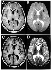
Cavitating Leukoencephalopathy in Fulminant Subacute Sclerosing Panencephalitis A 14-year-old girl presented with high-grade fever followed by recurrent episodes of generalized tonic–clonic seizures and altered sensorium, developing over a period of 2 weeks. She had a history of 2 to 3 episodes of generalized seizures and recurrent falls while walking, suggestive of negative myoclonus, 1 year previously for which she had been prescribed antiseizure medications, which were discontinued after 3 months. She continued to attend school, and no clear decline in scholastic performance was noted. She was an only child of non-consanguineous parents. She had history of a febrile exanthem at the age of 2 years 6 months, although measles was not confirmed serologically. She was admitted to another center in a mute and incontinent state at 2 weeks of symptom onset. Initial magnetic resonance imaging (MRI) of her brain showed bilateral temporal lobe hyperintensities (Fig.). Herpes encephalitis was suspected. Cerebrospinal fluid analysis showed 3 cells (lymphocytes), protein 26 mg/dL, and sugar 89.5 mg/dL. Cerebrospinal fluid (CSF) proved negative for herpes simplex virus, varicella-zoster, Japanese encephalitis, dengue, Epstein–Barr viruses, and autoimmune encephalitis panel. She was initiated on antiseizure medications. Injectable acyclovir 500 mg thrice daily was given for 3 weeks. Another possibility of seronegative autoimmune encephalitis was kept, and the patient was initiated on intravenous pulse methylprednisolone for 5 days. She continued to have persistent seizures and encephalopathy and was transferred to our center. At admission, the patient was intubated and mechanically ventilated for poor sensorium. A repeat MRI of her brain was performed at 2 months. Follow-up T2 and fluid-attenuated inversion recovery (FLAIR) axial images demonstrated multiple cystic lesions in bilateral cerebral hemispheres (arrows, Fig. ) and hyperintense grey and white matter (arrowheads). T1 axial contrast images (Fig.) demonstrated incomplete peripheral enhancement of the cysts (arrows) and nodular enhancement in thalami. Electroencephalogram (EEG) showed generalized delta slowing. Repeat review of her medical history revealed that the child was only partially vaccinated for measles. CSF analysis was repeated. It was acellular, with elevated protein (120 mg/dL) and normal sugar. CSF demonstrated raised anti-measles antibody titer in the CSF relative to the serum, enabling a diagnosis of subacute sclerosing panencephalitis (SSPE) based on the presence of clinical features (atypical), elevated CSF: serum anti-measles antibodies and elevated CSF protein, per modified Dyken’s criteria. CSF and serum (EIA) was performed. CSF/serum quotient was 11.6 (positive >1.5). Serum IgG measles 6,416 U/mL, CSF IgG measles 14,490 U/mL, serum total IgG 2,579 mg/ dL, and CSF total IgG 10.09 mg/dL (normal 0.3–4 mg/dL). The patient developed severe autonomic dysfunction soon thereafter and died of her illness within 2 months of the onset of symptoms. SSPE is a progressive disorder characterized by the presence of myoclonus, cognitive decline, and typical EEG changes. Our patient presented atypically in several ways – she lacked myoclonus, had an extremely rapid course, and displayed the development of cavitating leukoencephalopathy. Initial neuroimaging in SSPE may be
Journal Title: Annals of Neurology
Year Published: 2022
Link to full text (if available)
Share on Social Media: Sign Up to like & get
recommendations!