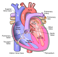
Photo from wikipedia
In this cadaver‐based study, we aimed to present a novel approach to pulmonary valve (PV) anatomy, morphometry, and geometry to offer comprehensive information on PV structure. The 182 autopsied human… Click to show full abstract
In this cadaver‐based study, we aimed to present a novel approach to pulmonary valve (PV) anatomy, morphometry, and geometry to offer comprehensive information on PV structure. The 182 autopsied human hearts were investigated morphometrically. The largest PV area was seen for the coaptation center plane, followed by basal ring and the tubular plane (626.7 ± 191.7 mm2 vs. 433.9 ± 133.6 mm2 vs. 290.0 ± 110.1 mm2, p < 0.001). In all leaflets, fenestrations are noted and occur in 12.5% of PVs. Only in 31.3% of PVs, the coaptation center is located in close vicinity of the PV geometric center. Similar‐sized sinuses were found in 35.7% of hearts, in the remaining cases, significant heterogeneity was seen in size. The mean sinus depth was: left anterior 15.59 ± 2.91 mm, posterior: 16.04 ± 2.82 mm and right anterior sinus: 16.21 ± 2.81 mm and the mean sinus height: left anterior 15.24 ± 3.10 mm, posterior: 19.12 ± 3.79 mm and right anterior sinus: 18.59 ± 4.03 mm. For males, the mean pulmonary root perimeters and areas were significantly larger than those for females. Multiple forward stepwise regression model showed that anthropometric variables might predict the coaptation center plane (sex, age, and heart weight; R2 = 33.8%), tubular plane (sex, age, and BSA; R2 = 20.5%) and basal ring level area (heart weight and sex; R2 = 17.1%). In conclusion, the largest pulmonary root area is observed at the coaptation center plane, followed by the basal ring and tubular plane. The PV geometric center usually does not overlap valve coaptation center. Significant heterogeneity is observed in the size of sinuses and leaflets within and between valves. Anthropometric variables may be used to predict pulmonary root dimensions.
Journal Title: Clinical Anatomy
Year Published: 2022
Link to full text (if available)
Share on Social Media: Sign Up to like & get
recommendations!