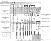
Photo from wikipedia
A small population of retinal ganglion cells expresses the photopigment melanopsin and function as autonomous photoreceptors. They encode global luminance levels critical for light‐mediated non‐image forming visual processes including circadian… Click to show full abstract
A small population of retinal ganglion cells expresses the photopigment melanopsin and function as autonomous photoreceptors. They encode global luminance levels critical for light‐mediated non‐image forming visual processes including circadian rhythms and the pupillary light reflex. There are five melanopsin ganglion cell subtypes (M1–M5). M1 and displaced M1 (M1d) cells have dendrites that ramify within the outermost layer of the inner plexiform layer. It was recently discovered that some melanopsin ganglion cells extend dendrites into the outer retina. Outer Retinal Dendrites (ORDs) either ramify within the outer plexiform layer (OPL) or the inner nuclear layer, and while present in the mature retina, are most abundant postnatally. Anatomical evidence for synaptic transmission between cone photoreceptor terminals and ORDs suggests a novel photoreceptor to ganglion cell connection in the mammalian retina. While it is known that the number of ORDs in the retina is developmentally regulated, little is known about the morphology, the cells from which they originate, or their spatial distribution throughout the retina. We analyzed the morphology of melanopsin‐immunopositive ORDs in the OPL at different developmental time points in the mouse retina and identified five types of ORDs originating from either M1 or M1d cells. However, a pattern emerges within these: ORDs from M1d cells are generally longer and more highly branched than ORDs from conventional M1 cells. Additionally, we found ORDs asymmetrically distributed to the dorsal retina. This morphological analysis provides the first step in identifying a potential role for biplexiform melanopsin ganglion cell ORDs.
Journal Title: Journal of Comparative Neurology
Year Published: 2017
Link to full text (if available)
Share on Social Media: Sign Up to like & get
recommendations!