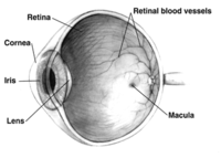
Photo from wikipedia
Loss of retinal ganglion cells (RGCs) underlies several forms of retinal disease including glaucomatous optic neuropathy, a leading cause of irreversible blindness. Several rare genetic disorders associated with cilia dysfunction… Click to show full abstract
Loss of retinal ganglion cells (RGCs) underlies several forms of retinal disease including glaucomatous optic neuropathy, a leading cause of irreversible blindness. Several rare genetic disorders associated with cilia dysfunction have retinal degeneration as a clinical hallmark. Much of the focus of ciliopathy associated blindness is on the connecting cilium of photoreceptors; however, RGCs also possess primary cilia. It is unclear what roles RGC cilia play, what proteins and signaling machinery localize to RGC cilia, or how RGC cilia are differentiated across the subtypes of RGCs. To better understand these questions, we assessed the presence or absence of a prototypical cilia marker Arl13b and a widely distributed neuronal cilia marker AC3 in different subtypes of mouse RGCs. Interestingly, not all RGC subtype cilia are the same and there are significant differences even among these standard cilia markers. Alpha‐RGCs positive for osteopontin, calretinin, and SMI32 primarily possess AC3‐positive cilia. Directionally selective RGCs that are CART positive or Trhr positive localize either Arl13b or AC3, respectively, in cilia. Intrinsically photosensitive RGCs differentially localize Arl13b and AC3 based on melanopsin expression. Taken together, we characterized the localization of gold standard cilia markers in different subtypes of RGCs and conclude that cilia within RGC subtypes may be differentially organized. Future studies aimed at understanding RGC cilia function will require a fundamental ability to observe the cilia across subtypes as their signaling protein composition is elucidated. A comprehensive understanding of RGC cilia may reveal opportunities to understanding how their dysfunction leads to retinal degeneration.
Journal Title: Journal of Comparative Neurology
Year Published: 2022
Link to full text (if available)
Share on Social Media: Sign Up to like & get
recommendations!