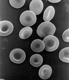
Photo from wikipedia
Iron deficiency independently predicts poor exercise capacity in heart failure with reduced ejection fraction (HFrEF), objectified as a low peak maximal oxygen consumption (VO2max). 1 Yet, precise mechanisms by which… Click to show full abstract
Iron deficiency independently predicts poor exercise capacity in heart failure with reduced ejection fraction (HFrEF), objectified as a low peak maximal oxygen consumption (VO2max). 1 Yet, precise mechanisms by which iron deficiency results in limited exercise capacity remain elusive. Peak VO2max is determined by the Fick equation as the difference between arterial O2 content (CaO2) and venous O2 content (CvO2) multiplied by cardiac output (CO) [VO2max= (CaO2 − CvO2)×CO]. Iron deficiency could impact all three components of the Fick equation thereby resulting in a blunted peak VO2max. However, the precise contribution of iron deficiency to each individual component of the Fick equation remains unknown. We prospectively included HFrEF patients admitted for haemodynamic guided decongestive therapy using a pulmonary artery catheter (PAC). All patients provided written informed consent and the study was approved by the institutional review board (Ziekenhuis Oost Limburg, Belgium). On the final day of the admission to the cardiac critical care unit a complete haemodynamic profile was registered at rest. Results of the laboratory analysis were used to stratify patients into an iron-deficient group (ferritin <100 μg/L or ferritin between 100 and 300 μg/L with a transferrin saturation <20%) and a non-iron-deficient group. Arterial O2 saturation (SaO2) and partial arterial O2 pressure (PaO2) were measured from an arterial blood sample obtained from a radial artery line. Mixed venous O2 saturation (SvO2) and partial venous O2 pressure (PvO2) were measured from a mixed venous sample from the PAC. CaO2 and CvO2 were calculated using the oxygen content formula [Ca/vO2 = (Hb× 1.36× Sa/vO2)+ (0.0031× Pa/vO2) with Hb denoting haemoglobin]. Cardiac output was monitored real-time using the automatic thermodilution application of the PAC (Swan-Ganz Continuous Cardiac Output Thermodilution Catheter 744HF75, Edwards Lifesciences, Irvine, CA, USA). Afterwards patients were asked to perform a symptom-limited supine bicycle exercise test (MOTOmed®Letto2, RECKTechnik GmbH&Co, Betzenweiler, Germany) under continuous haemodynamic monitoring. Patients were instructed to achieve 55–65 r.p.m. during eight cycles of 3 min at increasing workloads. At every increase of workload, an invasive haemodynamic assessment as well as arterial and mixed venous blood gases were obtained. Afterwards the impact of iron deficiency on contractile reserve (rise in CO and cardiac index from baseline to peak exercise) and peripheral O2 extraction (CaO2 –CvO2) between
Journal Title: European Journal of Heart Failure
Year Published: 2018
Link to full text (if available)
Share on Social Media: Sign Up to like & get
recommendations!