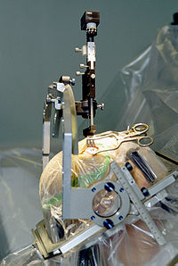
Photo from wikipedia
In patients with gliomas, changes in hemispheric specialization for language determined by magnetoencephalography (MEG) were analyzed to elucidate the impact of treatment and tumor recurrence on language networks. Demonstration of… Click to show full abstract
In patients with gliomas, changes in hemispheric specialization for language determined by magnetoencephalography (MEG) were analyzed to elucidate the impact of treatment and tumor recurrence on language networks. Demonstration of reorganization of language networks in these patients has significant implications on the prevention of postoperative functional loss and recovery. Whole‐brain activity during an auditory verb generation task was estimated from MEG recordings in a group of 73 patients with recurrent gliomas. Hemisphere of language dominance was estimated using the language laterality index (LI), a measure derived from the task. The initial scan was performed prior to resection; patients subsequently underwent surgery and adjuvant treatment. A second scan was performed upon recurrence prior to repeat resection. The relationship between the shift in LI between scans and demographics, anatomic location, pathology, and adjuvant treatment was analyzed. Laterality shifts were observed between scans; the median percent change was 29.1% across all patients. Laterality shift magnitude and relative direction were associated with the initial position of language dominance; patients with increased lateralization experienced greater shifts than those presenting more bilateral representation. A change in LI from left or right to bilateral (or vice versa) occurred in 23.3% of patients; complete switch occurred in 5.5% of patients. Patients with tumors within the language‐dominant hemisphere experienced significantly greater shifts than those with contralateral tumors. The majority of patients with glioma experience shifts in language network organization over time which correlate with the relative position of language lateralization and tumor location.
Journal Title: Human Brain Mapping
Year Published: 2019
Link to full text (if available)
Share on Social Media: Sign Up to like & get
recommendations!