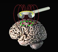
Photo from wikipedia
Individual neuroanatomy can influence motor responses to transcranial magnetic stimulation (TMS) and corticomotor excitability after intermittent theta burst stimulation (iTBS). The purpose of this study was to examine the relationship… Click to show full abstract
Individual neuroanatomy can influence motor responses to transcranial magnetic stimulation (TMS) and corticomotor excitability after intermittent theta burst stimulation (iTBS). The purpose of this study was to examine the relationship between individual neuroanatomy and both TMS response measured using resting motor threshold (RMT) and iTBS measured using motor evoked potentials (MEPs) targeting the biceps brachii and first dorsal interosseus (FDI). Ten nonimpaired individuals completed sham‐controlled iTBS sessions and underwent MRI, from which anatomically accurate head models were generated. Neuroanatomical parameters established through fiber tractography were fiber tract surface area (FTSA), tract fiber count (TFC), and brain scalp distance (BSD) at the point of stimulation. Cortical magnetic field induced electric field strength (EFS) was obtained using finite element simulations. A linear mixed effects model was used to assess effects of these parameters on RMT and iTBS (post‐iTBS MEPs). FDI RMT was dependent on interactions between EFS and both FTSA and TFC. Biceps RMT was dependent on interactions between EFS and and both FTSA and BSD. There was no groupwide effect of iTBS on the FDI but individual changes in corticomotor excitability scaled with RMT, EFS, BSD, and FTSA. iTBS targeting the biceps was facilitatory, and dependent on FTSA and TFC. MRI‐based measures of neuroanatomy highlight how individual anatomy affects motor system responses to different TMS paradigms and may be useful for selecting appropriate motor targets when designing TMS based therapies.
Journal Title: Human Brain Mapping
Year Published: 2022
Link to full text (if available)
Share on Social Media: Sign Up to like & get
recommendations!