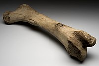
Photo from wikipedia
Individuals with type 2 diabetes mellitus (T2DM) have a greater risk of bone fracture compared with those with normal glucose tolerance (NGT). In contrast, individuals with impaired glucose tolerance (IGT)… Click to show full abstract
Individuals with type 2 diabetes mellitus (T2DM) have a greater risk of bone fracture compared with those with normal glucose tolerance (NGT). In contrast, individuals with impaired glucose tolerance (IGT) have a lower or similar risk of fracture. Our objective was to understand how progressive glycemic derangement affects advanced glycation endproduct (AGE) content, composition, and mechanical properties of iliac bone from postmenopausal women with NGT (n = 35, age = 65 ± 7 years, HbA1c = 5.8% ± 0.3%), IGT (n = 26, age = 64 ± 5 years, HbA1c = 6.0% ± 0.4%), and T2DM on insulin (n = 25, age = 64 ± 6 years, HbA1c = 9.1% ± 2.2%). AGEs were assessed in all samples using high‐performance liquid chromatography to measure pentosidine and in NGT/T2DM samples using multiphoton microscopy to spatially resolve the density of fluorescent AGEs (fAGEs). A subset of samples (n = 14 NGT, n = 14 T2DM) was analyzed with nanoindentation and Raman microscopy. Bone tissue from the T2DM group had greater concentrations of (i) pentosidine versus IGT (cortical +24%, p = 0.087; trabecular +35%, p = 0.007) and versus NGT (cortical +40%, p = 0.003; trabecular +35%, p = 0.004) and (ii) fAGE cross‐link density versus NGT (cortical +71%, p < 0.001; trabecular +44%, p < 0.001). Bone pentosidine content in the IGT group was lower than in the T2DM group and did not differ from the NGT group, indicating that the greater AGE content observed in T2DM occurs with progressive diabetes. Individuals with T2DM on metformin had lower cortical bone pentosidine compared with individuals not on metformin (−35%, p = 0.017). Cortical bone from the T2DM group was stiffer (+9%, p = 0.021) and harder (+8%, p = 0.039) versus the NGT group. Bone tissue AGEs, which embrittle bone, increased with worsening glycemic control assessed by HbA1c (Pen: R2 = 0.28, p < 0.001; fAGE density: R2 = 0.30, p < 0.001). These relationships suggest a potential mechanism by which bone fragility may increase despite greater tissue stiffness and hardness in individuals with T2DM; our results suggest that it occurs in the transition from IGT to overt T2DM. © 2022 American Society for Bone and Mineral Research (ASBMR).
Journal Title: Journal of Bone and Mineral Research
Year Published: 2022
Link to full text (if available)
Share on Social Media: Sign Up to like & get
recommendations!