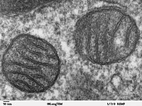
Photo from wikipedia
This study aimed to investigate the antitumor effect and the underlying molecular mechanism of eriodictyol on ovarian cancer cells. CaoV3 and A2780 were exposed to eriodictyol at different concentrations of 0-800… Click to show full abstract
This study aimed to investigate the antitumor effect and the underlying molecular mechanism of eriodictyol on ovarian cancer cells. CaoV3 and A2780 were exposed to eriodictyol at different concentrations of 0-800 μM. Cell apoptosis and viability were determined by TdT-mediated dUTP Nick-End Labeling (TUNEL) assay and Cell Counting Kit-8 (CCK-8) assay, respectively. Mitochondrial membrane potential was evaluated by flow cytometers with a JC-1 detection kit. Fe2+ content was evaluated using an iron assay kit. The section of tumor tissues was observed using hematoxylin-eosin (H&E) staining and nuclear factor erythroid 2-related factor 2 (Nrf2) expression was detected by immunohistochemistry (IHC) staining. Eriodictyol suppressed cell viability and induced cell apoptosis of CaoV3 and A2780 cells. Half maximal inhibitory concentration (IC50 ) value of CaoV3 at 24 and 48 h was (229.74 ± 5.13) μM and (38.44 ± 4.68) μM, and IC50 value of A2780 at 24 and 48 h was (248.32 ± 2.54) μM and (64.28 ± 3.19) μM. Fe2+ content and reactive oxygen species production were increased and protein levels of SLC7A11 and GPX4 were decreased by eriodictyol. Besides, eriodictyol reduced the ratio of JC-1 fluorescence ratio, glutathione and malondialdehyde contents but elevated Cytochrome C level. Nrf2 phosphorylation were obviously downregulated by eriodictyol. Finally, eriodictyol suppressed tumor growth, aggravated mitochondrial dysfunction and downregulated Nrf2 expression in tumor tissue in mice. Eriodictyol regulated ferroptosis, mitochondrial dysfunction and cell viability via Nrf2/HO-1/NQO1 signaling pathway in ovarian cancer.
Journal Title: Journal of biochemical and molecular toxicology
Year Published: 2023
Link to full text (if available)
Share on Social Media: Sign Up to like & get
recommendations!