Photo from wikipedia
Variceal bleeding is one of the major causes of death in patients affected by hepatic cirrhosis, together with hepatic failure and hepatocellular carcinoma (HCC). An early diagnosis and staging of… Click to show full abstract
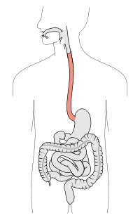
Variceal bleeding is one of the major causes of death in patients affected by hepatic cirrhosis, together with hepatic failure and hepatocellular carcinoma (HCC). An early diagnosis and staging of esophageal varices are therefore essential to provide effective treatment and prevent acute hemorrhage. Upper gastrointestinal endoscopy is undoubtedly the gold standard for diagnosis of varices, as it not only can reliably demonstrate the presence and grade of varices, but it also allows endoscopic treatment when necessary. Nevertheless, endoscopy remains an invasive and uncomfortable procedure for patients. The procedure is also costly, and serious complications such us perforation and hemorrhage can occur, however rarely. In addition, the risk of variceal development in early and compensated cirrhosis is low, and many endoscopies might be
Journal Title: Journal of Clinical Ultrasound
Year Published: 2022
Link to full text (if available)
Share on Social Media: Sign Up to like & get
recommendations!