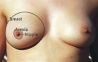
Photo from wikipedia
Worldwide, breast cancer (BrCa) is currently the leading cause of deaths associated to malignant lesions in adult women. Given that some studies have mentioned that peritumoral adipocytes may contribute to… Click to show full abstract
Worldwide, breast cancer (BrCa) is currently the leading cause of deaths associated to malignant lesions in adult women. Given that some studies have mentioned that peritumoral adipocytes may contribute to breast carcinogenesis, present work sought to quantitative evaluate the morphometry of these cells in a group of adult women. Three thousand six hundred sixty four breast adipocytes, that came from biopsies of a group of adult females with different types of breast carcinomas (ductal, lobular, and mixed) and one with normal tissues, were evaluated through an image analysis (IA) process regarding six morphometric descriptors: area (A), perimeter (P), Feret diameter (FD), aspect ratio (AR), roundness factor (RF), and fractal dimension of cellular contour (FDC). Data showed that the adipocytes of the normal tissues group were bigger (A: 3398 ± 2331 µm2, P: 239 ± 83 µm, and FD: 79.9 ± 24.5 µm) than those from BrCa samples (A: 2860 ± 1933 µm2, P: 214 ± 66 µm, and FD: 73.2 ± 22.5 µm), and presented a more irregular contour (FDC of 1.370 ± 0.037 for normal group and of 1.335 ± 0.049 for the oncologic one). Moreover, it could be accounted that adipocytes of mixed carcinomas were largest (FD: 75.1 ± 22.4 µm) than those of lobular lesions (FD: 61.6 ± 22.6 µm), while the adipocytes of ductal carcinomas were the most oval (AR: 1.421 ± 0.524) and roughest (FDC: 1.324 ± 0.050) cells. IA results suggest that BrCa lesions can be categorized through a quantitative morphometric evaluation of peritumoral adipocytes. These findings could let the development of an analytical tool to help the Pathologist to enhance the accuracy of the oncologic diagnose.
Journal Title: Microscopy Research and Technique
Year Published: 2018
Link to full text (if available)
Share on Social Media: Sign Up to like & get
recommendations!