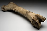
Photo from wikipedia
Bone is an organic matrix containing type I collagen (40%), mineral crystals (45%), and water (15%). The water that exists in the cortical bone can be classified as bound water… Click to show full abstract
Bone is an organic matrix containing type I collagen (40%), mineral crystals (45%), and water (15%). The water that exists in the cortical bone can be classified as bound water (BW) and free or pore water (PW) representing water binding to the organic matrix and residing in Haversian canals and lacunae-canalicular systems, respectively. The T2* value is less than 20 μs for the collagen backbone protons, between 300 and 400 μs for the BW, and more than 1 ms for the PW in the cortical bone in 3 T MRI. Therefore, it is essential to characterize the relaxation of the BW and PW for evaluating the individual contribution of the BW and PW to the mechanical properties of the cortical bone using ultrashort echo time (UTE) or short echo time (STE) because of the short T2 value of the cortical bone. Prior research has shown that the relaxations of the BW and PW have opposite correlations with biomechanical measures of bone competence. The content and MR relaxation of the total water (TW), BW, and PW are sensitive to the structural and mechanical properties of the cortical bone. Thus, measure of these characteristics provides a noninvasive method of evaluating the quality of the cortical bone. For example, Bae et al. used UTE technique to measure the content and T2* of water and reported that the TW content and PW content are positively correlated with porosity of cortical bone but negatively correlated with the failure strain and ultimate stress. They also found that the long T2* fraction (PW) is positively correlated with, while the short T2* fraction and the short T2* (BW) are negatively correlated with the porosity of cortical bone. Furthermore, the failure strain was positively correlated with the short T2* value, and the failure energy was positively correlated with both short and long T2* values. In addition to the T2* relaxation, the T1 relaxation of the BW and the PW in the cortical bone has also been investigated. For instance, Chen et al. employed 3D UTE imaging to measure the T1 relaxation of bovine cortical bone, showing a T1 value of 208, 545, and 131 ms for TW, PW, and BW, respectively. Using UTE technique, Jerban et al showed that the T1 value does not correlate with bone microstructural and mechanical properties. However, the correlation between T1 value of BW and PW with the biomechanical measures of bone competence has not been well investigated using clinical MR scanners. In the article entitled “Cortical Bone Mechanical Assessment via Free Water Relaxometry at 3T”, the authors quantified free water (or PW as mentioned) content of cortical bone in 20 tibiae of freshly slaughtered cows using STE (TE, 1.3 ms) pulse sequences, including inversion recovery (IR), variable flip angle (VFA), and variable repetition time (VTR) methods, to quantify T1 values of tibial cortical bone and obtained indirect information about bone microstructure and mechanical properties. They also investigated the contribution of the T1 value for the PW in the cortical bone to the mechanical properties of the cortical bone via mechanical compression test. In T1 measurements, their results showed that the T1 values measured by the VTR and VFA methods (VTR-T1 and VFA-T1, respectively) did not differ from that measured by the IR method (IR-T1). In mechanical properties, their results disclosed that the toughness was negatively correlated with IR-T1 (r = 0.77, P = 0.01) and that the toughness (r = 0.68, P = 0.00), ultimate stress (r = 0.71, P = 0.00), and yield stress (r = 0.62, P = 0.00) were negatively correlated with the VFA-T1, while none of the mechanical parameters correlated with the VTR-T1. Accordingly, the authors concluded that VFA-T1 outperforms VTR-T1 in evaluating the relationship between the PW and the mechanical properties of the cortical bone using STE at a 3 T MR scanner. It is interesting and clinically important to evaluate the mechanical properties of cortical bone to reflect the potential risk evaluation of bone fracture. Overall speaking, it is a very good idea to measure the T1 by three STE pulse sequences (IR, VTR, and VFA) so that the authors have the
Journal Title: Journal of Magnetic Resonance Imaging
Year Published: 2021
Link to full text (if available)
Share on Social Media: Sign Up to like & get
recommendations!