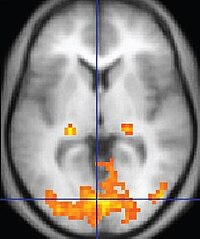
Photo from wikipedia
Localized regions of left–right image intensity asymmetry (LRIA) were incidentally observed on T2‐weighted (T2‐w) and T1‐weighted (T1‐w) diagnostic magnetic resonance imaging (MRI) images. Suspicion of herpes encephalitis resulted in unnecessary… Click to show full abstract
Localized regions of left–right image intensity asymmetry (LRIA) were incidentally observed on T2‐weighted (T2‐w) and T1‐weighted (T1‐w) diagnostic magnetic resonance imaging (MRI) images. Suspicion of herpes encephalitis resulted in unnecessary follow‐up imaging. A nonbiological imaging artifact that can lead to diagnostic uncertainty was identified.
Journal Title: Journal of Magnetic Resonance Imaging
Year Published: 2022
Link to full text (if available)
Share on Social Media: Sign Up to like & get
recommendations!