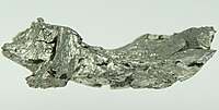
Photo from wikipedia
Currently, the gadolinium retention in the brain after the use of contrast agents is studied by T1‐weighted magnetic resonance imaging (MRI) (T1w) and T1 mapping. The former does not provide… Click to show full abstract
Currently, the gadolinium retention in the brain after the use of contrast agents is studied by T1‐weighted magnetic resonance imaging (MRI) (T1w) and T1 mapping. The former does not provide easily quantifiable data and the latter requires prolonged scanning and is sensitive to motion. T2 mapping may provide an alternative approach. Animal studies of gadolinium retention are complicated by repeated intravenous (IV) dosing, whereas intraperitoneal (IP) injections might be sufficient.
Journal Title: Journal of Magnetic Resonance Imaging
Year Published: 2022
Link to full text (if available)
Share on Social Media: Sign Up to like & get
recommendations!