Photo from wikipedia
We illustrate the intravascular ultrasound (US) findings in the evaluation of left gonadal vein anatomic variations. During a 2‐year period, 4 consecutive patients (mean age, 37 years; range, 28–45 years)… Click to show full abstract
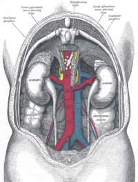
We illustrate the intravascular ultrasound (US) findings in the evaluation of left gonadal vein anatomic variations. During a 2‐year period, 4 consecutive patients (mean age, 37 years; range, 28–45 years) with left‐sided varicocele underwent embolization. Intravascular US examinations and retrograde venography were performed to assess varicocele anatomy. Anatomic variants were recorded and categorized. A comparison between intravascular US and fluoroscopic findings was performed. The Fisher exact test was used for statistical analysis (P < .05). Technical success was achieved in all cases. There was a statistically significant difference in the maximum gonadal vein diameter between venography and intravascular US (P = .0087). Intravascular US showed left gonadal vein anatomic variations and better ability in the evaluation of the vein diameter.
Journal Title: Journal of Ultrasound in Medicine
Year Published: 2019
Link to full text (if available)
Share on Social Media: Sign Up to like & get
recommendations!