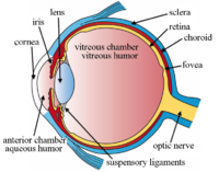
Photo from wikipedia
We read with interest the article by Chen et al. [1] describing choroidal remodeling in eyes with polypoidal choroidal vasculopathy (PCV) following treatment with verteporfin photodynamic therapy (PDT). Using spectraldomain… Click to show full abstract
We read with interest the article by Chen et al. [1] describing choroidal remodeling in eyes with polypoidal choroidal vasculopathy (PCV) following treatment with verteporfin photodynamic therapy (PDT). Using spectraldomain optical coherence tomography (OCT), the authors reported early increases in central foveal thickness, subretinal fluid height, and choroidal thickness after PDT. At a subsequent review performed between 1 to 3months, the above parameters were reduced compared to baseline [1]. It is known that following treatment with PDT, subretinal fluid, or retinal edema may increase. This is believed to result from an inflammatory response and upregulation of vascular endothelial growth factor (VEGF). Importantly, this may result in worsening of visual acuity, which could be alarming to the patient. It would be interesting to know the change in visual acuity in this cohort, and whether this correlated with the changes in retinal or choroidal parameters. Another important consideration is that patients in the current study were treated with full-dose PDT. However, for PCV, it is more common to perform PDT together with intravitreal injections of anti-VEGF agents [2–4]. The concurrent use of anti-VEGF agents may lessen the inflammatory response and up-regulation of VEGF induced by PDT, and may mitigate the transient increase in subretinal fluid and choroidal permeability. For example, in a multicentre randomized controlled clinical trial (EVEREST II study) [2], central retinal thickness was reduced compared to baseline 1 month after treatment with PDT and intravitreal ranibizumab. It would be interesting to perform similar imaging studies on a cohort of patients treated with combination therapy to observe whether there are any differences in choroidal remodeling compared to the current group treated with PDT monotherapy. The authors reported that polyps manifested with hypoflow on OCT angiography (OCTA) [1]. It is important to point out, however, that not all polypoidal lesions appear as regions of hypoflow.Theappearance of polypoidal lesions on OCTA are quite variable, with between 36.2% to 55.3% of polyps detectable using OCTA [5,6]. Currently, the gold standard for diagnosis of PCV is indocyanine green angiography, which allows visualization of the full extent of the PCV lesion in order to plan PDT treatment [7–9]. In summary, the current study adds to our knowledge of the effects of PDT treatment on the choroid and retina. It would be interesting to observe the changes following treatment with combination PDT and intravitreal antiVEGFagents, as this is amore common treatment for PCV.
Journal Title: Lasers in Surgery and Medicine
Year Published: 2018
Link to full text (if available)
Share on Social Media: Sign Up to like & get
recommendations!