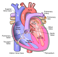
Photo from wikipedia
A 50-year old female with acute-on-chronic liver failure and MELD score of 34 was referred for LDLT. Her husband volunteered to be donor. Computed tomography (CT) revealed a Type III… Click to show full abstract
A 50-year old female with acute-on-chronic liver failure and MELD score of 34 was referred for LDLT. Her husband volunteered to be donor. Computed tomography (CT) revealed a Type III right portal vein (2) (RPPV) and isolated right posterior hepatic artery (RPHA). The right anterior section was drained predominantly by the large RHV that received two prominent V5 branches(orange arrow, Figure 1a) instead of the fine middle hepatic vein (MHV). Segment 7 was drained by two V7s while segment 6 was drained by the inferior right hepatic vein (IRHV). Biliary tree anatomy showed both S6 and S7 ducts drained into right posterior duct (RPD).
Journal Title: Liver Transplantation
Year Published: 2020
Link to full text (if available)
Share on Social Media: Sign Up to like & get
recommendations!