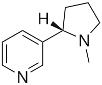
Photo from wikipedia
We thank M. Jubeau and J. Gondin for their interest in our work (1). We are grateful that they have pointed our attention to their recent work on neuromuscular electrical… Click to show full abstract
We thank M. Jubeau and J. Gondin for their interest in our work (1). We are grateful that they have pointed our attention to their recent work on neuromuscular electrical stimulation (NMES) (2), which we unintentionally missed during the preparation of our manuscript (1). Both studies, however, differ on crucial points. In contrast to the nonlocalized volume-selection utilizing coil sensitivity, as used in our study, they applied a chemical shift image sequence with a better defined volume selection at the expense of temporal resolution (43.5 s (2) vs. 5 s (1)), which induces substantial temporal averaging over shifting metabolite peaks. Further differences exist with regard to the investigated muscle and stimulation pattern. Nevertheless, our interpretation that the identified pH compartments correspond to different fiber types was supported by their observation of pH-heterogeneity in spectra of homogeneous muscle regions with outer volume contaminations between 9% to 10%. The main difference to the work of Jubeau et al. and the novelty of our study is that we maximized the NMESinduced pH-heterogeneity by a short and strong stimulation (PCr depletion was completed after the first 50 s (1) vs. 400 s in (2)), which would likewise explain the substantially higher shifts of the acidic pH-component (pH< 6.4) in our study. Thus, spectroscopic series were acquired with the best possible time resolution (5 s) while accepting compromises with regard to signal localization and signal-to-noise ratio (SNR). Under these conditions, we observed a split of Pi into three Pi components in all volunteers, which thus far only was detected for voluntary muscle contractions (3). Because it can be assumed that proton enrichment by glycolytic activity and proton exchange are competitive processes, the lower stimulation intensity in the approach of Jubeau et al. may be responsible for smaller pH differences and for the absence of an additional split. Their presentation of the vastus lateralis spectra during NMES stimulation (middle spectrum in the bottom block Figure 1 in Jubeau et al. (2)) lets us assume that the linewidth of the Pi-1 component is larger than for Pi-2, possibly indicating a superposition of two nonresolved components. Unfortunately, we did not find indications of linewidths in their paper. Due to the imprecisely defined muscular contributors to the spectra in our study, it can be assumed that the high pH component represents a contamination of signal from the soleus, which already was discussed in our paper (1). As illustrated by the changes in T2*-w images between preand postfatigue (Supporting Figs. S9-S12 in Stutzig et al. (1)), these changes are limited to the medial gastrocnemius muscle, and thus no pH-changes are expected in the soleus and the lateral gastrocnemius, whereas all dynamic series apart from one indicate at least a small acidic shift compared to the resting pH (Fig. 7 in Stutzig et al. (1)). We assume that individual differences are responsible for the prominent shift of two volunteers showing pH of 6.3 and 6.4 because methodical errors or different training status of the subjects can be excluded as an explanation. The SNR of PCr in the individual resting spectra was between 13 and 21 (16.2 6 2.8) without any averaging process, which was renounced because the Pi components were distinguishable from signal-free regions of the spectra and indicated a continuous temporal evolution of the pH shifts. In summary, we believe that the exercise procedure and intensity substantially affect the metabolic response and that the investigation of different load conditions helps to understand the underlying physiological processes. Although our work is restricted by some methodical limitations, we nevertheless think that our results indicate that high-intensity NMES stimulation is suitable to generate pH-heterogeneity between different compartments in the stimulated muscle, which can be detected by P-MRS. Furthermore, we agree with Jubeau and Gondin that higher magnetic field and localized acquisition techniques would provide more relevant information about the phenomenon of the Pi splitting.
Journal Title: Magnetic Resonance in Medicine
Year Published: 2017
Link to full text (if available)
Share on Social Media: Sign Up to like & get
recommendations!