Photo from wikipedia
Sir Peter Mansfield, who died on February 8, 2017, was the co-inventor of MRI and a pioneer of solid-state NMR. His origination of MRI and subsequent development of many of… Click to show full abstract
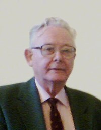
Sir Peter Mansfield, who died on February 8, 2017, was the co-inventor of MRI and a pioneer of solid-state NMR. His origination of MRI and subsequent development of many of the key ideas that still underpin this remarkable, noninvasive imaging technique was most prominently recognized through the award of the Nobel Prize in Physiology or Medicine in 2003—an award that he shared with Paul Lauterbur (1). Sir Peter was born and educated in Lambeth in inner London. He left school at the age of 15 with no formal qualifications, to take up the role as a printer’s assistant. An interest in rocketry, partially stemming from his exposure to V2 rocket attacks on London during World War II, subsequently secured him a job in the rocket-propulsion department of Britain’s Ministry of Supply. By taking evening classes while working on rockets during the day, he acquired the qualifications needed to study for a degree in physics, joining Queen Mary University in London in 1954. Sir Peter’s final-year undergraduate project involved building a transistor-based continuous-wave NMR spectrometer, and success in this enterprise brought Sir Peter to the attention of Jack Powles (one of the earliest users of NMR in the United Kingdom), who offered him a PhD studentship. During his doctoral studies, Sir Peter built a pulsed spectrometer for solid-state NMR experiments, and using this system he discovered the solid echo in which the effects of dipolar couplings are partially reversed by application of a pair of 90 pulses (2). On completion of his PhD, Sir Peter took up a postdoctoral position with Charles Slichter at the University of Illinois, where he worked on NMR studies of doped metals. While working in Urbana, Sir Peter was recruited to a lectureship in Nottingham University’s Department of Physics by Raymond Andrew. Taking up that appointment in 1964, Sir Peter stayed at Nottingham University for the rest of his career, despite being courted by many other academic institutions. In Nottingham, he initially worked on multiple pulse sequences for line-narrowing in solid-state NMR (3). To implement this work, Sir Peter constructed one of the first computercontrolled NMR spectrometers. This was based around a Honeywell computer (Morris Plains, NJ, USA) with just 4 kB of memory to accommodate the data, and a machine code implementation of the fast Fourier transform. These developments led him to think about ways of measuring crystal structure in solids via NMR “diffraction,” and the realization that magnetic field gradients were needed to make this possible. His first one-dimensional NMR imaging experiments were carried out on a small sample consisting of multiple, millimeter-thick plates of camphor (a waxy solid), and these were reported in an article in the Journal of Physics in 1973 (4), the same year that Paul Lauterbur published his seminal letter to Nature on NMR “zeugmatography” (5). Quite quickly, the focus of Sir Peter’s research moved almost completely onto NMR imaging, and at the 18th Ampere Congress, which was held in Nottingham in 1974 and also attended by Paul Lauterbur, new results were reported in what must have been one of the first-ever conference sessions devoted to “NMR image formation.” Over the next few years, Sir Peter introduced the sliceselection process (6), and produced the first images of live human anatomy (7) (the finger of Andrew Maudsley, who was Sir Peter’s PhD student at the time). This image helped to convince the United Kingdom’s Medical Research Council to provide the funding needed for Sir Peter to construct a first 0.1T whole-body scanner (8). Despite concerns at the time about the possibility that the voltages induced in the torso by switching on and off magnetic field gradients during the imaging process might cause a cardiac arrest (9), Sir Peter volunteered for the first experiments (a re-enactment was recorded later for the United Kingdom’s “Tomorrow’s World” television program), and the results were reported a few days later at the 1978 Experimental Nuclear Magnetic Resonance Conference. Around this time, Sir Peter also conceived the echo-planar imaging (EPI) technique (10), which allows a 2D image to be generated from a single free induction decay. Variants of EPI are of course used today in almost all MRI-based functional brain imaging experiments, but it is worth remembering that at the time of its invention, standard MR image acquisition times were of the order of tens of minutes. It took more than 10 years of work by Sir Peter (11), including the invention of actively screened gradient coils (12), now used in all MRI scanners, and continued innovation in the use of cutting edge computer technology, to bring EPI to the point where it could be adopted for widespread use. This typifies the determination that he showed throughout his career: His ideas often reflected what the underlying physics suggested would be possible rather than what the current technology allowed. However, he was always prepared to persevere with the development of the techniques and hardware needed to allow him to eventually realize his vision. In the early 1990s, Sir Peter built the first 500-MHz NMR microscope (13), the first 3T human scanner (14), and he carried out early studies of porous media using MRI (15). Sir Peter joined the editorial board of Magnetic Resonance in Medicine when the journal was first established in 1984, and the journal is still proud to name him as an honorary member of the editorial board, along with Richard Ernst, Paul Lauterbur, and Erwin Hahn. Much of Sir Peter’s later work was published in MRM. For example, he published four papers in the journal’s first volume, including the influential paper that first described EPIbased chemical shift imaging (16). Sir Peter was elected to the United Kingdom’s Royal Society in 1987, knighted in 1993, and won many honors and prizes in addition to Received 3 July 2017; accepted 6 July 2017 DOI 10.1002/mrm.26855 Published online 00 Month 2017 in Wiley Online Library (wileyonlinelibrary. com). Magnetic Resonance in Medicine 00:000–000 (2017)
Journal Title: Magnetic resonance in medicine
Year Published: 2017
Link to full text (if available)
Share on Social Media: Sign Up to like & get
recommendations!