Photo from wikipedia
The abdominal cavity is an enclosed entity and consequently, intra-abdominal pressure (Pabd) persistently engages all intra-abdominal organs, including the bladder and the proximal two-thirds of the urethra. This is true… Click to show full abstract
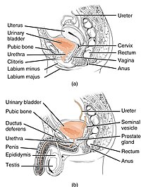
The abdominal cavity is an enclosed entity and consequently, intra-abdominal pressure (Pabd) persistently engages all intra-abdominal organs, including the bladder and the proximal two-thirds of the urethra. This is true for both continent and incontinent women, both at rest and during stress (Fig. 1). The urethra and the anterior vaginal wall are conjoined by the endopelvic fascia and by other paraurethral connective tissues wherein the posterior pubourethral ligaments (PUL) are of fundamental importance. The urethra is thus part of the anterior vaginal wall and the pelvic floor. When the pelvic floor is pressed downward during stress, the urethra ‘‘follows.’’ This is devastating for the urethral sphincter mechanism in cases where the bladder neck does not descend to a similar extent. This is the situation for women with stress urinary incontinence (SUI). Instead of being stopped by a backboard effect, the proximal urethra is stopped and funneled by hanging on the bladder neck. This forced funneling of the inner urethra is due to a failure of the suburethral supporting structures, mainly the PUL, to maintain the normal spatial relationship between the urethra and bladder neck, which is necessary to prevent the proximal urethra from reaching a hanging position. As such, an abnormal spatial relationship displays as an enlarged posterior urethrovesical angle. The forced funneling of the collapsed inner urethra overwhelms the high urethral opening pressure. A miniscule meatus internus is a perfect seal. This is analogous to blowing up a rubber balloon when the initiation of expansion may be nearly impossible. Pascal’s principle of fluid mechanics postulates that when the radius (r) of the meatus internus widens, the outflow-distending force (Fd) increases abruptly, which is described as follows in Pascal’s formula: Fd1⁄4 (aLPPþ Pdet) pi r2; (aLPP1⁄4 abdominal leak point pressure, Pdet1⁄4detrusor pressure). Additionally the length of the functional urethra is shortened in proportion to the depth of the funnel. The urethral high-pressure zone can consequently be engaged. In the cases of a short functional urethral length and/or a low urethral closure pressure at rest (UCP), the urethral resistance is low and facilitates urethral opening and urine leakage. Thus, the forced funneling exerts one external shearing force (Fs) and enhances one internal distending force (Fd). The proximal two-thirds of the urethra, inclusive of the highpressure zone, persist inside the abdominal cavity and accordingly the aLPP is transmitted. However, when the proximal urethra is funneled, the shearing force Fs and the distending force Fd counterbalance its manifestation. At aLPP, the urethra opens and leaks; UCPþ aLPP Fs Fd1⁄40. Hypermobile SUI is when the bladder neck is hypermobile and the proximal urethra at stress descends further than the bladder neck. Hypomobile SUI is when the bladder neck is hypomobile and the proximal urethra at stress descends further than the bladder neck. In hypomobile SUI, the urethral downward distance to reach a hanging position is short; in severe cases, the urethra hangs on the bladder neck and displays funneling even at rest. Regardless of hypermobile or hypomobile SUI, the UCP can be low or high, which has minor impact (see the above formulas). A collapsed inner urethra with a low UCP withstands very high valsalva pressures. Conversely, urethral funneling has fundamental significance. If the urethral funnel radius increases from 0.5 to 5mm, the outflow distending force (Fd) increases 100 times. The inner urethra is the ‘‘flow-controlling zone’’ and the midurethral high-pressure zone/UCP is of relatively lesser significance. The PUL are broadly attached to the paraurethral tissues at the junction of the upper one-third and distal two-thirds of the urethra. This junction, which corresponds to a vaginal point (v.p.), is likely the optimal position for the tension-free vaginal tape (TVT) to enforce a correct spatial relationship at stress between the proximal urethra and bladder neck. Similarly, the v.p. is also the midpoint of the intra-abdominal urethra. The distance between the v.p. at rest and the v.p. at aLPP is the ‘‘therapeutic window’’ (t.w.). A TVT located inside the t.w. is curative. Nevertheless, the tape at rest should be set accurately and tension-free to not incriminate the t.w. ‘‘safety margin.’’ In hypermobile SUI, the t.w. is large. In hypomobile SUI the t.w. is small and the correct placement of the TVT is restricted, which
Journal Title: Neurourology and Urodynamics
Year Published: 2017
Link to full text (if available)
Share on Social Media: Sign Up to like & get
recommendations!