Photo from wikipedia
Fluorescent imaging, especially in living tissue, has become a key method in modern life sciences, with the development of new tools for sample preparation, imaging, and data analysis continuously advancing… Click to show full abstract
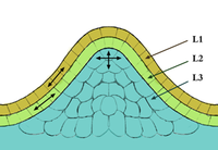
Fluorescent imaging, especially in living tissue, has become a key method in modern life sciences, with the development of new tools for sample preparation, imaging, and data analysis continuously advancing our understanding of biological principles. Here, we present our strategy for in vivo imaging of the Arabidopsis shoot apical meristem (SAM), a central structure in plant development. We implement simplifications to previously published workflows and present a novel approach to subsequentially image the meristem from multiple angles at high resolution. This tool may represent a valuable resource for shoot meristem-centered research in general, but also for studies on plasmodesmata or intercellular connectivity within the SAM: via the analysis of fluorescently labeled plasmodesmata-localized proteins, via the tracing of fluorescent dyes, via analyzing the cell-to-cell mobility of fluorescently labeled proteins, but also via the analysis of morphological features of meristematic cells in mutants or upon perturbation of symplastic connectivity.
Journal Title: Methods in molecular biology
Year Published: 2022
Link to full text (if available)
Share on Social Media: Sign Up to like & get
recommendations!