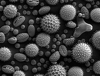
Photo from wikipedia
Quantification of plasmodesmata density on cell interfaces of plant tissues, particularly of leaves, has been a long-standing challenge. Using electron microscopy alone to quantify plasmodesmata is difficult because of the… Click to show full abstract
Quantification of plasmodesmata density on cell interfaces of plant tissues, particularly of leaves, has been a long-standing challenge. Using electron microscopy alone to quantify plasmodesmata is difficult because of the limited surface area coverage per image and hence the need to examine large numbers of sections for robust quantification. Fluorescence microscopy provides the larger surface area coverage per image but can only visualize pit fields and not individual plasmodesma. Moreover, in pigmented tissue like leaves, imaging cell interfaces beyond the epidermal layer would also require accurate sectioning. The advent of tissue clearing techniques such as PEA-CLARITY provided the opportunity to capture all pit fields within the leaf without resorting to sectioning. This paved the way toward the development of a more robust and precise plasmodesmata density quantification method by combining the three-dimensional immunolocalization fluorescence microscopy with scanning electron microscopy (SEM). Here, I describe a protocol to quantify plasmodesmata density on cell interfaces between mesophyll and bundle sheath in C3 and C4 monocot leaves.
Journal Title: Methods in molecular biology
Year Published: 2022
Link to full text (if available)
Share on Social Media: Sign Up to like & get
recommendations!