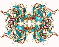
Photo from wikipedia
Immunofluorescence microscopy is an invaluable tool for the study of biological processes at the cellular level. While the localization of surface-exposed antigens can easily be determined using fluorescent antibodies, localization… Click to show full abstract
Immunofluorescence microscopy is an invaluable tool for the study of biological processes at the cellular level. While the localization of surface-exposed antigens can easily be determined using fluorescent antibodies, localization of intracellular antigens requires permeabilization of the bacterial cell wall and membrane. Here, we describe an immunofluorescence protocol tailored specifically for Streptococcus pyogenes, applying the phage lysin PlyC for cell wall permeabilization. This protocol allows a high level of morphological preservation, suitable for high-resolution microscopy. With slight modification, this protocol could also be used for other Gram-positive pathogens.
Journal Title: Methods in molecular biology
Year Published: 2017
Link to full text (if available)
Share on Social Media: Sign Up to like & get
recommendations!