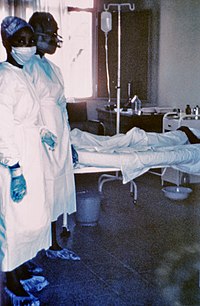
Photo from wikipedia
Ebola virus (EBOV) replicates in host cells, where both viral and cellular components show morphological changes during the process of viral replication from entry to budding. These steps in the… Click to show full abstract
Ebola virus (EBOV) replicates in host cells, where both viral and cellular components show morphological changes during the process of viral replication from entry to budding. These steps in the replication cycle can be studied using electron microscopy (EM), including transmission electron microscopy (TEM) and scanning electron microscopy (SEM), which is one of the most useful methods for visualizing EBOV particles and EBOV-infected cells at the ultrastructural level. This chapter describes conventional methods for EM sample preparation of cultured cells infected with EBOV.
Journal Title: Methods in molecular biology
Year Published: 2017
Link to full text (if available)
Share on Social Media: Sign Up to like & get
recommendations!