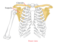
Photo from wikipedia
Purpose Aim of this study is to establish an objective and easily applicable method that will allow clinicians to quantitatively assess scapular dyskinesis during clinical examination using a computer tablet… Click to show full abstract
Purpose Aim of this study is to establish an objective and easily applicable method that will allow clinicians to quantitatively assess scapular dyskinesis during clinical examination using a computer tablet software. Hypothesis is that dyskinetic scapulae present greater motion—deviation from the thoracic wall—compared to the non-dyskinetic ones and that the software will be able to record those differences. Methods Twenty-five patients and 19 healthy individuals were clinically evaluated for the presence of dyskinesis or not. According to the clinical diagnosis, the observations were divided into three groups; A. Dyskinetic scapulae with symptoms ( n = 25), B. Contralateral non-dyskinetic scapulae without symptoms ( n = 25), C. Healthy control scapulae ( n = 38). Then, all individuals were tested using a tablet with the PIVOT™ image-based analysis software (PIVOT, Impellia, Pittsburgh, PA, USA). The motion produced by the scapula medial border and inferior angle deviation from the thoracic wall was recorded. Results The deviation of the medial border and inferior angle of the scapula from the thoracic wall was 24.6 ± 7.3 mm in Group A, 14.7 ± 4.9 mm in Group B, and 12.4 ± 5.2 mm in Group C. The motion recorded in the dyskinetic scapulae group was significantly greater than both the contralateral non-dyskinetic scapulae group ( p < 0.01) and the healthy control scapulae group ( p < 0.01). Conclusion The PIVOT™ software was efficient to detect significant differences in the motion between dyskinetic and non-dyskinetic scapulae. This system can support the clinical diagnosis of dyskinesis with a numeric value, which not only contributes to scapula dyskinesis grading but also to the evaluation of the progress and efficacy of the applied treatment, thus providing a feedback to the clinician and the patient. Level of evidence IV, laboratory study.
Journal Title: Knee Surgery, Sports Traumatology, Arthroscopy
Year Published: 2020
Link to full text (if available)
Share on Social Media: Sign Up to like & get
recommendations!