Photo from wikipedia
Purpose To define the length-change patterns of the superficial medial collateral ligament (sMCL), deep MCL (dMCL), and posterior oblique ligament (POL) across knee flexion and with applied anterior and rotational… Click to show full abstract
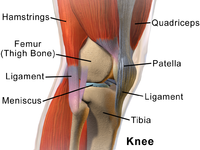
Purpose To define the length-change patterns of the superficial medial collateral ligament (sMCL), deep MCL (dMCL), and posterior oblique ligament (POL) across knee flexion and with applied anterior and rotational loads, and to relate these findings to their functions in knee stability and to surgical repair or reconstruction. Methods Ten cadaveric knees were mounted in a kinematics rig with loaded quadriceps, ITB, and hamstrings. Length changes of the anterior and posterior fibres of the sMCL, dMCL, and POL were recorded from 0° to 100° flexion by use of a linear displacement transducer and normalised to lengths at 0° flexion. Measurements were repeated with no external load, 90 N anterior draw force, and 5 Nm internal and 5 Nm external rotation torque applied. Results The anterior sMCL lengthened with flexion ( p < 0.01) and further lengthened by external rotation ( p < 0.001). The posterior sMCL slackened with flexion ( p < 0.001), but was lengthened by internal rotation ( p < 0.05). External rotation lengthened the anterior dMCL fibres by 10% throughout flexion ( p < 0.001). sMCL release allowed the dMCL to become taut with valgus rotation ( p < 0.001). The anterior and posterior POL fibres slackened with flexion ( p < 0.001), but were elongated by internal rotation ( p < 0.001). Conclusion The structures of the medial ligament complex react differently to knee flexion and applied loads. Structures attaching posterior to the medial epicondyle are taut in extension, whereas the anterior sMCL, attaching anterior to the epicondyle, is tensioned during flexion. The anterior dMCL is elongated by external rotation. These data offer the basis for MCL repair and reconstruction techniques regarding graft positioning and tensioning.
Journal Title: Knee Surgery, Sports Traumatology, Arthroscopy
Year Published: 2020
Link to full text (if available)
Share on Social Media: Sign Up to like & get
recommendations!