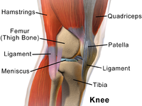
Photo from wikipedia
The purpose of this study is to examine the association between the development of articular cartilage pathology and knee rotation after single-bundle anterior cruciate ligament (ACL) reconstruction. Seventeen patients that… Click to show full abstract
The purpose of this study is to examine the association between the development of articular cartilage pathology and knee rotation after single-bundle anterior cruciate ligament (ACL) reconstruction. Seventeen patients that underwent single-bundle ACL reconstruction and did not have any cartilage lesions at the time of surgery based on the Outerbridge classification or meniscal injury that required meniscectomy > 20% were examined by MRI and in the biomechanics laboratory at a 6-year minimum follow-up. Cartilage lesions that occurred after reconstruction were graded on MRI according to a modified Noyes scale. For cartilage evaluation, the lateral and medial femoral condyles were divided into 9 segments each (lateral, central, and medial third and each third was divided into anterior, central, and posterior segment). Tibial rotation during a pivoting task was measured with optoelectronic motion analysis system and side-to-side differences of tibial rotation between the reconstructed and contralateral intact knees were calculated. The association between the total modified Noyes scale score (outcome variable) and side-to-side differences of tibial rotation after controlling for meniscectomy and meniscal repair was investigated with hierarchical regression models. Side-to-side difference of tibial rotation was associated with total modified Noyes scale score (p = 0.015, β = 0.667, adjusted R2 = 42.1%). All patients developed new cartilage lesions in MRI located mainly at the central region of the lateral femoral condyle and less frequently in the central and anterior regions of the medial femoral condyle. Abnormally increased tibial rotation that persists after ACL-R is significantly associated with the development of new articular cartilage lesions at mean 8.4 years after reconstruction which were located mainly at the central region of the LFC and secondarily in the central and anterior regions of the MFC (more superficial lesions). These findings suggest that there is emerging evidence that abnormal rotational kinematics is a potential risk factor for the pathogenesis and onset of posttraumatic articular cartilage degeneration after ACLR. IV.
Journal Title: Knee Surgery, Sports Traumatology, Arthroscopy
Year Published: 2021
Link to full text (if available)
Share on Social Media: Sign Up to like & get
recommendations!