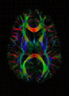
Photo from wikipedia
Neurolymphoma (NL) is a rare entity representing peripheral nervous system infiltration bymalignant lymphocytes [1]. It is most commonly seen in the context of aggressive sub-types of lymphoma, such as diffuse… Click to show full abstract
Neurolymphoma (NL) is a rare entity representing peripheral nervous system infiltration bymalignant lymphocytes [1]. It is most commonly seen in the context of aggressive sub-types of lymphoma, such as diffuse large B cell non-Hodgkin’s lymphoma [2]. Its exact incidence is not well documented [3]. While only 20% of patients with NL have evidence of systemic involvement at the time of staging, systemic disease is commonly reported at autopsy [4]. Involvement of peripheral nerves without systemic manifestations, i.e. primary neurolymphomatosis, has been most commonly reported within the sciatic nerve [5]. Clinical presentation is variable and includes cranial neuropathy, painful and painless mononeuropathy, painful radiculopathy, and polyneuropathy [3]. The diagnosis of NL hinges upon both radiological and histopathological examination, in view of its rarity and diverse clinical presentation. NL most frequently involves the proximal peripheral nerves and brachial and lumbosacral plexuses. Imaging appearances range from diffuse enlargement of the peripheral nerve to a mass-like appearance of the involved nerve [2]. NL demonstrates T2 hyperintensity and a nodular pattern or fusiform enlargement [4]. T2-weighted imaging usually demonstrates scattered areas of hypointensity within the mass in keeping with displaced nerve fascicles within the tumor. MRI features that suggest a diagnosis of NL include enlargement of nerves or nerve roots beyond the root sleeve [1]. Post-contrast imaging may show homogenous or heterogeneous enhancemen t o f t he mas s l e s i on [5 ] . Ultrasonographic features of NL are very different from peripheral neurogenic tumors. In NL, sonographic features include infiltration of the nerve by a surrounding mass, focal thickening of the nerve or of individual fascicles within the nerve. Nerve biopsies may yield false-negative results due to the patchy distribution of NL [4]. The differential considerations include peripheral spread of malignant disease. Perineural spread ofmalignancymay occur in head and neck tumors, gastrointestinal malignancy, breast cancer, prostate cancer, and cervical cancer. Other differential considerations of NL include inflammatory neuropathy. At MR imaging, inflammatory neuritis is depicted as diffuse thickening of the affected nerves and returns a high T2 signal. Peripheral nerve sheath tumors can mimic NL, however, the former appear heterogenous at MR imaging. Peripheral nerve sheath tumors return a low signal intensity on T1-weighted imaging and intermediate to high signal on T2-weighted imaging [4]. Differential diagnoses in patients with a prior history of lymphoma includes lymphoma-associated vasculitis and amyloidosis [4]. In our case, MRI showed a fusiform soft tissue mass extending from the inferior portion of the right upper arm and crossing the elbow joint (Fig. 1a, b). The mass measured 16 cm in craniocaudal dimension with a maximum crosssectional dimension of 4.6 × 3 cm. The mass appeared to arise from themedian nerve, which itself was noted to be thickened. The mass interdigitated with the adjacent nerve fibers (Fig. 1b, c) and displaced the adjacent brachial artery. It demonstrated intermediate to low signal intensity (SI) to adjacent muscle on T1 (Fig. 1a) and high SI on T2 (Fig. 1b) and fat-suppressed imaging (Fig. 1c). Multiple dot-like dark signal foci were noted on proton density and fat-suppressed T2-weighted imaging (Fig. 1c) consistent with displaced nerve fascicles in the tumor. These foci were depicted as low signal striations along the course of the nerve on coronal and sagittal imaging (Fig. 1b). The T2 hyperintensity and enlargement of the nerves were possible indicators of malignancy [4]. The case presentation can be found at https://doi.org/10.1007/s00256018-3125-z
Journal Title: Skeletal Radiology
Year Published: 2018
Link to full text (if available)
Share on Social Media: Sign Up to like & get
recommendations!