Photo from wikipedia
ObjectiveThe purpose of our study was to determine the main anatomical features of the calcarine sulcus using a 3-T MRI.MethodsFifty human brains have been explored using an MRI 3-T in… Click to show full abstract
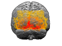
ObjectiveThe purpose of our study was to determine the main anatomical features of the calcarine sulcus using a 3-T MRI.MethodsFifty human brains have been explored using an MRI 3-T in Doctors Center in Beirut (Lebanon).ResultsThe calcarine sulcus was identified in 100% of cases. In most cases, it had a continuous aspect with several peaks. In all our specimens, the calcarine sulcus crosses the parieto-occipital fissure. The majority of their collateral branches and their connections with other sulci were located at the level of the calcarine sulcus properly. In the majority of specimens, the deepest part of the anterior calcarine sulcus forms a protrusion in the occipital horn of the lateral ventricle called calcar avis.ConclusionOur study emphasizes the fact that the course patterns of the calcarine sulcus are highly variable. The description of the main anatomical features of the calcarine sulcus obtained from our study can be used as a reference for fMRI exploration and is useful for brain surgery.
Journal Title: Surgical and Radiologic Anatomy
Year Published: 2018
Link to full text (if available)
Share on Social Media: Sign Up to like & get
recommendations!