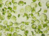
Photo from wikipedia
Protoplasts of six cabbage accessions were isolated from leaf mesophyll and cultured in the presence of 0.01, 0.1 and 1.0 µM phytosulfokine–α (PSK-α) and in a PSK-free control medium. PSK-α was… Click to show full abstract
Protoplasts of six cabbage accessions were isolated from leaf mesophyll and cultured in the presence of 0.01, 0.1 and 1.0 µM phytosulfokine–α (PSK-α) and in a PSK-free control medium. PSK-α was applied for 10 days and later, protoplast-derived cells were cultured in the PSK-free medium. Supplementation of the culture medium with PSK-α showed a dose-dependent effect on the mitotic activity of cultured cells. On the 15th day of culture, the highest mitotic activity of protoplast-derived cells was observed in cultures treated with 0.1 µM of PSK-α, and ranged from 14 to 60% dependent on the accession. The number of multi-cell structures was also higher (90–93%) on this medium compared to the control (77–80%). Analysis of cellulose regeneration in cultured protoplasts after Calcofluor White staining showed that this process was not synchronous, but depended instead on the presence of PSK-α in the culture medium, and was more pronounced in the low-responding accession. Sustained cell divisions led to formation of microcallus colonies, subjected to regeneration on solid media. Supplementation of the regeneration media with 0.1 µM of PSK significantly increased shoot regeneration compared to the control media. Moreover, enhanced regeneration was observed from calluses developed from cells treated with PSK-α at the early stages of development and later transferred for regeneration onto the media supplemented with 0.1 µM of this peptide.
Journal Title: Journal of Plant Growth Regulation
Year Published: 2018
Link to full text (if available)
Share on Social Media: Sign Up to like & get
recommendations!