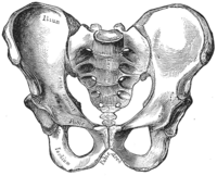
Photo from wikipedia
The levator ani (LA) complex in high-type imperforate anus (H-IA), low-type imperforate anus (L-IA), and Hirschsprung’s disease (HD) patients as controls were documented using magnetic resonance imaging (MRI) and compared… Click to show full abstract
The levator ani (LA) complex in high-type imperforate anus (H-IA), low-type imperforate anus (L-IA), and Hirschsprung’s disease (HD) patients as controls were documented using magnetic resonance imaging (MRI) and compared for symmetry. Mean left:right LA thickness ratio (LA ratio), and deviation of the LA from the pubococcygeal line (PCL; LA angle) were calculated from thin-slice MRI images (axial 2 mm, coronal 2 mm, and sagittal 3 mm) of the puborectalis and pubococcygeus taken parallel to the PCL under sedation in H-IA (n=14), L-IA (n=16), and HD (n=9). MRI scans were performed between January 2018 and June 2021. LA were significantly thinner in H-IA (1.78±0.46 mm) compared with L-IA (2.97±0.55 mm) and controls (2.87±0.32 mm), p<0.0001. LA ratio was significantly lower in H-IA (0.71±0.15) compared with L-IA (0.93±0.04), and controls (0.91±0.06), p<0.0001. Mean LA-angle was significantly different in H-IA, 10.8° (range 6°–19°), versus L-IA and controls, both zero degrees (range 0°–5°), p<0.0001, respectively. LA was confirmed to be significantly asymmetric in H-IA. Because outcome of surgical repair involving a midline incision, such as posterior sagittal anorectoplasty could be impaired, pediatric surgeons are advised to plan surgical intervention for H-IA carefully and appropriately.
Journal Title: Pediatric Surgery International
Year Published: 2022
Link to full text (if available)
Share on Social Media: Sign Up to like & get
recommendations!