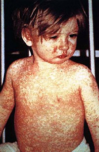
Photo from wikipedia
Skin color is determined by the number of melanin granules produced by melanocytes that are transferred to keratinocytes. Melanin synthesis and the distribution of melanosomes to keratinocytes within the epidermal… Click to show full abstract
Skin color is determined by the number of melanin granules produced by melanocytes that are transferred to keratinocytes. Melanin synthesis and the distribution of melanosomes to keratinocytes within the epidermal melanin unit (EMU) within the skin of vitiligo patients have been poorly studied. The ultrastructure and distribution of melanosomes in melanocytes and surrounding keratinocytes in perilesional vitiligo and normal skin were investigated using transmission electron microscopy (TEM). Furthermore, we performed a quantitative analysis of melanosome distribution within the EMUs with scatter plot. Melanosome count within keratinocytes increased significantly compared with melanocytes in perilesional stable vitiligo (P < 0.001), perilesional halo nevi (P < 0.01) and the controls (P < 0.01), but not in perilesional active vitiligo. Furthermore, melanosome counts within melanocytes and their surrounding keratinocytes in perilesional active vitiligo skin decreased significantly compared with the other groups. In addition, taking the means—standard error of melanosome count within melanocytes and keratinocytes in healthy controls as a normal lower limit, EMUs were graded into 3 stages (I–III). Perilesional active vitiligo presented a significantly different constitution in stages compared to other groups (P < 0.001). The distribution and constitution of melanosomes were normal in halo nevi. Impaired melanin synthesis and melanosome transfer are involved in the pathogenesis of vitiligo. Active vitiligo varies in stages and in stage II, EMUs are slightly impaired, but can be resuscitated, providing a golden opportunity with the potential to achieve desired repigmentation with an appropriate therapeutic choice. Adverse milieu may also contribute to the low melanosome count in keratinocytes.
Journal Title: Archives of Dermatological Research
Year Published: 2017
Link to full text (if available)
Share on Social Media: Sign Up to like & get
recommendations!