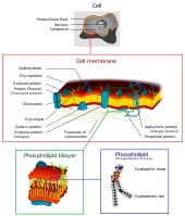
Photo from wikipedia
Human amniotic membrane (HAM) is traditionally used for the treatment of non-healing wounds. However, high density of HAM-matrix (HAM-M) diminishes cellular contribution for successful tissue regeneration. Herein we investigated whether… Click to show full abstract
Human amniotic membrane (HAM) is traditionally used for the treatment of non-healing wounds. However, high density of HAM-matrix (HAM-M) diminishes cellular contribution for successful tissue regeneration. Herein we investigated whether a bioengineered micro-porous three-dimensional (3D) HAM-scaffold (HAM-S) could promote healing in ischemic wounds in diabetic type 1 rat. HAM-S was prepared from freshly decellularized HAM. Then, 30 days after inducing diabetes, an ischemic circular excision was generated on rats’ skin. The diabetic animals were randomly divided into untreated (Diabetic group), engrafted with HAM-M (D-HAM-M group) and HAM-S (D-HAM-S group). Also, non-diabeticuntreated rats (Healthy group) were considered as control. Stereological, molecular, and tensiometrical assessments were performed on post-surgical days 7, 14, and 21. We found that the volumes of new epidermis and dermis, the numerical density of epidermal basal cells and fibroblasts, the length density of blood vessels, the numbers of proliferating cells and collagen deposition as well as biomechanical properties of healed wound were significantly higher in D-HAM-S group in most cases compared those of the diabetic group, or even in some cases compared to D-HAM-M group. Furthermore, in D-HAM-S group, the transcripts for genes contributing to regeneration (Tgf-β, bFgf and Vegf) upregulated more than those of D-HAM-M group, when compared to diabetic ones. Overall, the HAM-S had more impact on delayed wound healing process compared to traditional use of intact HAM. It is therefore suggested that the bioengineered three dimensional micro-porous HAM-S is more suitable for cells adhesion, penetration, and migration for contributing to wounded tissue regeneration.
Journal Title: Archives of Dermatological Research
Year Published: 2020
Link to full text (if available)
Share on Social Media: Sign Up to like & get
recommendations!