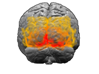
Photo from wikipedia
Spontaneous nystagmus (SN) in dorsal medullary infarction is usually horizontal or mixed horizontal-torsional with a small vertical component [1]. The slow phases of SN may be directed toward or away… Click to show full abstract
Spontaneous nystagmus (SN) in dorsal medullary infarction is usually horizontal or mixed horizontal-torsional with a small vertical component [1]. The slow phases of SN may be directed toward or away from the side of the lesion according to the involved neural structure. The horizontal SN with a torsional component beating away from the side of the lesion side may similar with that in peripheral vestibulopathy reflecting involvement of the vestibular nuclei. On the other hand, damages in the nucleus prepositus hypoglossi or inferior cerebellar peduncle (ICP) result in ipsilesional SN [2]. In patients with a central lesion mainly involving the posterior fossa, the SN was not typically attenuated by visual fixation, paradoxically SN can be even accentuated during visual fixation [3]. However, in dorsolateral medullary infarction, SN can be attenuated in amplitude under visual fixation similar with that in peripheral vestibulopathy [2]. Vertical nystagmus rarely converts to the opposite direction during convergence [4], but there has been no report on the reversal of the horizontal component of spontaneous nystagmus (SN) during visual fixation. We experienced a patient with dorsal medullary infarction who showed a reversal of the horizontal component of SN during visual fixation. A 55-year-old man developed a sudden onset of prolonged vertigo and imbalance on the day before admission. On admission, the patient showed a left beating SN in bedside examination (i.e., in the light). He had right beating SN when the cover of VNG goggle covered patient’s eyes during video-nystagmography (VNG) test with a progressive slow drift of eyeball to the right side (i.e., ocular lateropulsion) and a relocation of eye to the central position. The right beating SN was augmented after head-shaking. During visual fixation in the VNG test, the horizontal component of SN was converted to the left side (i.e., a reversal of SN) (Video 1). SN was purely horizontal, so we could not check the reverse of vertical or torsional component of SN. The slow phase velocities of SN without and with fixation were both linear shape and maximal slow phase velocity of SN without fixation was faster than that under fixation (10 deg/s vs. 6 deg/s, respectively). The amplitude of SN under visual fixation was decreased by the time and only lasted for 10–12 s and disappeared (Fig. 1a). Horizontal gaze-evoked nystagmus was not evident. Horizontal saccade test showed saccadic hypometria to the left side and saccadic movements was interrupted by left-beating nystagmus. Horizontal smooth pursuit eye movement toward the left side was impaired. The patient had limb ataxia on the right side. He could stand without support but veered to the right when attempting to walk. He had no skew deviation or ocular torsion. Ocular and cervical vestibular-evoked myogenic potentials, subjective visual vertical, video head-impulse, bithermal caloric and rotatory chair tests were all normal. Diffusion-weighted images revealed an acute infarct selectively involving the caudal dorsal medulla on the right side (Fig. 1b). After 3 weeks, the right beating SN was persistent during without fixation and the intensity was attenuated during fixation, but its direction was not converted. Our patient with dorsal medullary infarction showed a unique feature of SN in which, the horizontal component of SN during darkness was reversed its direction under visual fixation. In the dorsal medulla, anatomical structures responsible for ipsilesional SN are the ICP or nucleus prepositus Electronic supplementary material The online version of this article (https ://doi.org/10.1007/s0041 5-020-09754 -y) contains supplementary material, which is available to authorized users.
Journal Title: Journal of Neurology
Year Published: 2020
Link to full text (if available)
Share on Social Media: Sign Up to like & get
recommendations!