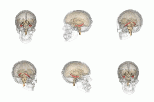
Photo from wikipedia
Neurofeedback training may improve cognitive function in patients with neurological disorders. However, the underlying cerebral mechanisms of such improvements are poorly understood. Therefore, we aimed to investigate MRI correlates of… Click to show full abstract
Neurofeedback training may improve cognitive function in patients with neurological disorders. However, the underlying cerebral mechanisms of such improvements are poorly understood. Therefore, we aimed to investigate MRI correlates of cognitive improvement after EEG-based neurofeedback training in patients with MS (pwMS). Fourteen pwMS underwent ten neurofeedback training sessions within 3–4 weeks at home using a tele-rehabilitation system. Half of the pwMS (N = 7, responders) learned to self-regulate sensorimotor rhythm (SMR, 12–15 Hz) by visual feedback and improved cognitively after training, whereas the remainder (non-responders, n = 7) did not. Diffusion-tensor imaging and resting-state fMRI of the brain was performed before and after training. We analyzed fractional anisotropy (FA) and functional connectivity (FC) of the default-mode, sensorimotor (SMN) and salience network (SAL). At baseline, responders and non-responders were comparable regarding sex, age, education, disease duration, physical and cognitive impairment, and MRI parameters. After training, compared to non-responders, responders showed increased FA and FC within the SAL and SMN. Cognitive improvement correlated with increased FC in SAL and a correlation trend with increased FA was observed. This exploratory study suggests that successful neurofeedback training may not only lead to cognitive improvement, but also to increases in brain microstructure and functional connectivity.
Journal Title: Journal of Neurology
Year Published: 2021
Link to full text (if available)
Share on Social Media: Sign Up to like & get
recommendations!