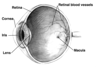
Photo from wikipedia
Background/aimsThe purpose of the study was to evaluate the retinal and choroidal changes via optical coherence tomography angiography (OCTA) in patients who received hydroxychloroquine (HCQ).MethodsSixty eyes of 60 female patients… Click to show full abstract
Background/aimsThe purpose of the study was to evaluate the retinal and choroidal changes via optical coherence tomography angiography (OCTA) in patients who received hydroxychloroquine (HCQ).MethodsSixty eyes of 60 female patients who received HCQ were included in the study. Patients were categorized into two groups as high-risk (≥ 5 years) and low-risk (< 5 years) in terms of HCQ-induced retinal toxicity. Spectral domain-OCT, OCTA, and visual field tests were performed. Retinal thickness, vascular density, flow rates, choroidal thickness (CT), and visual field parameters were compared between the groups, and the correlation between total HCQ cumulative dose, duration of use, and these parameters was assessed.ResultsCompared to low-risk group, patients in the high-risk group had vascular density loss (p < 0.05). In this group, foveal avascular zone (FAZ) was found to be wider (p < 0.05). Retinal and choroidal flow rates were found to be decreased markedly in the high-risk group (p < 0.05). CT was found to be thinner in the high-risk group (p < 0.05). HCQ cumulative dose and duration of use had a negative significant correlation with all vascular density, flow rate, CT parameters, and positive significant correlation with FAZ parameters (p < 0.05). In visual field tests, mean defect (MD) was found to be increased in the high-risk group (p < 0.05). Moreover, MD had a positive correlation with HCQ cumulative dose and duration of use (p < 0.05).ConclusionsEvaluation of microvascular changes via OCTA may contribute to the early detection of HCQ-induced retinal toxicity, which cannot be detected through other imaging devices, at the stage when it is reversible.
Journal Title: Graefe's Archive for Clinical and Experimental Ophthalmology
Year Published: 2018
Link to full text (if available)
Share on Social Media: Sign Up to like & get
recommendations!