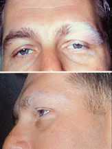
Photo from wikipedia
To prospectively evaluate the dynamic changes of the full-field electroretinogram (ff-ERG) and its association with inflammatory signs in patients with Vogt-Koyanagi-Harada disease (VKHD) followed up after acute onset. Twelve acute… Click to show full abstract
To prospectively evaluate the dynamic changes of the full-field electroretinogram (ff-ERG) and its association with inflammatory signs in patients with Vogt-Koyanagi-Harada disease (VKHD) followed up after acute onset. Twelve acute VKHD patients, who were followed up for at least 24 months, were enrolled at a tertiary center from June 2011 to January 2017. Treatment consisted of intravenous methylprednisolone followed by 1 mg/kg/day of oral prednisone with a slow tapering associated with late non-steroidal immunosuppressive therapy in previously defined cases. Inflammation was systematically evaluated with clinical and posterior segment imaging (PSI) exams (fluorescein angiography, FA, indocyanine green angiography, ICGA, enhanced depth imaging optical coherence tomography, EDI-OCT). A ff-ERG was performed upon enrollment as well as at predefined intervals. Scotopic ff-ERG parameters changes between the 12th and 24th months defined the ERG-stable or ERG-worsening groups. “Flare” was defined as an appearance or worsening of inflammatory signs (after the initial 6 months following disease onset) under the predefined treatment protocol. ff-ERG parameters initially improved in all eyes; in the evaluation between the 12th and 24th months, ff-ERG results were stable in 17 eyes (71 %) and worsened in 7 eyes (29 %). Subnormal ff-ERG results were observed in 15 eyes (62 %) at the 24th month. On the other hand, the flare was observed in 8 eyes (33 %) as cells in the anterior chamber and in 24 eyes (100 %) as any PSI inflammatory sign. The ERG-worsening group presented thicker subfoveal choroid at the first month (p = 0.001) and fluctuations in choroidal thickness more often during follow-up when compared to the ERG-stable group (p = 0.02). Scotopic ff-ERG parameters worsened between the 12th and 24th months in a quarter of the patients. Subclinical inflammation detected as an increase in CT seems to be related to worsening in visual function measured with ffERG.
Journal Title: Graefe's Archive for Clinical and Experimental Ophthalmology
Year Published: 2019
Link to full text (if available)
Share on Social Media: Sign Up to like & get
recommendations!