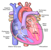
Photo from wikipedia
Lymphangiomas are comprised of aggregates of lymphatic vessels, considered to represent either aberrant embryogenic remnants or developing secondary to obstruction. Lymphangiomas primary to the heart and pericardial are exceedingly rare,… Click to show full abstract
Lymphangiomas are comprised of aggregates of lymphatic vessels, considered to represent either aberrant embryogenic remnants or developing secondary to obstruction. Lymphangiomas primary to the heart and pericardial are exceedingly rare, and to date sparingly reported in individual case reports. In this study, the histopathologic, clinical, and radiologic features of 35 cases of cardiac/pericardial lymphangiomas described in the literature to date together with four cases from our own institution (39 cases in total) are examined to provide clinicopathologic characterization. Cardiac/pericardial lymphangiomas were identified in both children and adults, with two cases initially discovered in utero. If presenting with symptoms, patients most commonly exhibited respiratory distress/dyspnea. By X-ray, a widened cardiac silhouette could be noted, and echocardiogram generally showed an echogenic mass with cystic and septal components. On computed tomography (CT) and magnetic resonance imaging (MRI), cystic and septal components were again observed, with CT showing an absence of calcifications or macroscopic fat. Most lymphangiomas were pericardial (specifically visceral) based, and frequently situated in the right atrioventricular groove. A majority of cases proceeded to surgical resection, with no evidence of recurrence post-operatively. Grossly, lesions had a median size of 6 cm and in almost all cases were multicystic/multilocular. Microscopically, the lymphangiomas were composed of lymphatic spaces lined by endothelial cells that specifically express podoplanin (D2-40) with immunoperoxidase staining. Further investigation with a larger and more uniformly organized cohort is required to better characterize the clinicopathologic features of lymphangiomas of this unusual anatomic location.
Journal Title: Virchows Archiv
Year Published: 2022
Link to full text (if available)
Share on Social Media: Sign Up to like & get
recommendations!