Photo from wikipedia
Background We investigated the understudied anatomical variations of the superior petrosal vein (SPV) complex (SPVC), which may play some role in dictating the individual complication risk following SPVC injury. Methods… Click to show full abstract
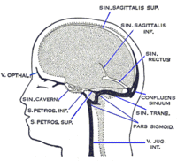
Background We investigated the understudied anatomical variations of the superior petrosal vein (SPV) complex (SPVC), which may play some role in dictating the individual complication risk following SPVC injury. Methods Microvascular decompressions of the trigeminal nerve between September 2012 and July 2016. All operations utilized an SPVC preserving technique. Preoperative balanced fast field echo (bFFE) magnetic resonance imaging, or equivalent sequences, and operative videos were studied for individual SPVC anatomical features. Results Applied imaging and operative SPVC anatomy were described for fifty patients (mean age, 67.18 years; female sex and right-sided operations, 58% each). An SPVC component was sacrificed intentionally in 6 and unintentionally in only 7 cases. Twenty-nine different individual variations were observed; 80% of SPVCs had either 2 SPVs with 3 or 1 SPV with 2, 3, or 4 direct tributaries. Most SPVCs had 1 SPV (64%) and 2 SPVs (32%). The SPV drainage point into the superior petrosal sinus was predominantly between the internal auditory meatus and Meckel cave (85.7% of cases). The vein of the cerebellopontine fissure was the most frequent direct tributary (86%), followed by the pontotrigeminal vein in 80% of SPVCs. Petrosal-galenic anastomosis was detected in at least 38% of cases. At least 1 SPV in 54% of the cases and at least 1 direct tributary in 90% disturbed the operative field. The tributaries were more commonly sacrificed. Conclusions The extensive anatomical variation of SPVC is depicted. Most SPVCs fall into 4 common general configurations and can usually be preserved. BFFE or equivalent sequences remarkably facilitated the intraoperative understanding of the individual SPVC in most cases.
Journal Title: Acta Neurochirurgica
Year Published: 2019
Link to full text (if available)
Share on Social Media: Sign Up to like & get
recommendations!