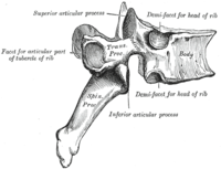
Photo from wikipedia
Background Magnetic resonance imaging (MRI) is important in the assessment of degenerative spine disease. However, its role is limited in the identification of spinal instability; therefore, weight-bearing and dynamic studies… Click to show full abstract
Background Magnetic resonance imaging (MRI) is important in the assessment of degenerative spine disease. However, its role is limited in the identification of spinal instability; therefore, weight-bearing and dynamic studies like X-rays are required. The supine position eliminates the gravitational pull, corrects the vertebral slippage, and opens the facet joints leading to the collection of the synovial fluid into the joint space, which is detected on the MRI and can serve as a marker for instability. We aim to compare the facet fluid, facet hypertrophy, facet angle, and disc degenerative changes among the patients presenting with degenerative spondylolisthesis (DS) and those without. Methods We performed a retrospective review for all the patients treated at our institution from January 2015 to December 2016. Facet Fluid Index (FFI) (ratio of facet fluid width and facet joint width) was calculated to assess the joint fluid. The percentage of spondylolisthesis was measured on X-rays. Each radiological parameter was compared between the two groups, i.e., patients with DS and patients without DS. A p value < 0.05 was considered significant. Results In total, 61 patients, 28 with DS and 33 without DS, were enrolled. Baseline characteristics were similar in the two groups ( p > 0.05). The average values of FFI, facet fluid width, and the difference between the superior and inferior facet were significantly higher in the group with instability ( p < 0.05). Multivariate analysis demonstrated a 4.44 (95% confidence interval [CI] 2.03–5.365) times increase in the odds of instability with a unit increase in FFI, p < 0.0001. Conclusions We report a positive linear correlation between the facet joint effusion and facet hypertrophy on MRI and the percentage of vertebral translation on X-ray. Prospective studies will determine if these markers can play a role in predicting spinal instability.
Journal Title: Acta Neurochirurgica
Year Published: 2021
Link to full text (if available)
Share on Social Media: Sign Up to like & get
recommendations!