Photo from wikipedia
In the article ‘Beyond magnification and illumination: Preliminary clinical experience with the 4K 3D ORBEYETM exoscope and a literature review’, Amoo and his colleagues introduce readers to their own experience… Click to show full abstract
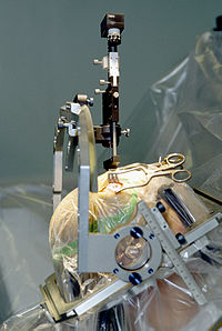
In the article ‘Beyond magnification and illumination: Preliminary clinical experience with the 4K 3D ORBEYETM exoscope and a literature review’, Amoo and his colleagues introduce readers to their own experience with the ORBEYETM exoscope (Olympus, Tokyo, Japan) in a series of 18 neurosurgical procedures. This device enables a highresolution, three-dimensional (3D) magnified and illuminated intraoperative visualization of the surgical field on a 55-inch monitor using special 3D glasses. Contrary to ‘traditional’ endoscopic systems that provide two-dimensional (2D) images on the monitor, the exoscope might serve as an alternative to the ‘classic’ operating microscope (OM) with stereoscopic visual and illuminational characteristics. Thus, Amoo et al. focused on the aspects of intraoperative visualization and on the proposed possible ergonomic advances compared to the OM. Besides evaluation of their own experience, they reviewed the current literature of the use of different 4K 3D exoscopes in the field of neurosurgery. The two following aspects are important: first, the technical development of the OM and, second, the technical development of the (neuro)endoscope since the beginning of the twentieth century. Despite the remarkable technical development of neurosurgical armamentarium over the past decades including novel optic systems, so far, the OM is one of the most important tools. It enables ‘direct’ stereoscopic visualization. The ENT surgeon Carl Olof Nylén (1892–1978) was the first physician, who applied a (monocular) surgical microscope in the operation room at the University of Stockholm in 1921 [4, 5]. One year later, the University’s head of the department Gunnar Holmgren (1875–1954) used a binocular operating microscope attached with a light source [4, 5]. Although being used by an increasing number of ENT surgeons in the following years, it took more than 30 years to introduce the microscope into the neurosurgical operating room. In 1957, Theodore Kurze removed a neurilemmoma from cranial nerve VII in a 5-year-old patient [4, 8], and in 1965, the operative microscope was used for intracranial aneurysm surgery by the surgeons J. Lawrence Pool and Robert P. Colton [4, 9]. Today, neurosurgery is the leading field in using the operating microscope and its additional elements such as highdefinition (HD) monitors, fluorescence imaging and imageguided surgical features [4]. Since the invention of Zeiss OPMI 1 in 1953, constant improvements regarding handling of the OM and technical applications were developed [4, 8]. Besides technical improvements for better illumination, ranges of magnification and longer working distance and changes concerning stability, flexibility, share of view and ergonomic and manoeuvrability were made. One important aspect is the share of view of the surgeon, the surgeon’s assistant and the scrub nurse by the attachment of a second eyepiece and a screen respectively. In general, modern OM are equipped with a selection of different binoculars for rotation in different heights and positions, full range of movement and several ranges of tilt of the optics carrier. Further, the newer generations’ OM has This article is part of the Topical Collection on Neurosurgery general
Journal Title: Acta Neurochirurgica
Year Published: 2021
Link to full text (if available)
Share on Social Media: Sign Up to like & get
recommendations!