Photo from wikipedia
Trigeminal neuralgia is the most common example of craniofacial neuralgia. Its etiology is unknown and is characterized by severe episodes of paroxysmal pain. The trigeminal ganglion and its adjacent anatomical… Click to show full abstract
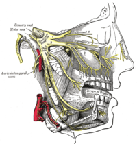
Trigeminal neuralgia is the most common example of craniofacial neuralgia. Its etiology is unknown and is characterized by severe episodes of paroxysmal pain. The trigeminal ganglion and its adjacent anatomical structures have a complex anatomy. The foramen ovale is of great importance during surgical procedures such as percutaneous trigeminal rhizotomy for trigeminal neuralgia. We aimed to identify the anatomical structures associated with the trigeminal ganglion and radiofrequency rhizotomy on cadavers and investigate their relationship with the electrodes used during rhizotomy to determine the contribution of the electrode diameter and length to the effectiveness of the lesion formation on the ganglion. Five fresh-frozen cadaver heads injected with red silicone/latex were used. A percutaneous puncture was made by inserting of a cannula through the foramen ovale to create a pathway for electrodes. The relationships between the electrodes, Meckel’s cave, trigeminal ganglion, and neurovascular structures were observed and morphometric measurements were obtained using a digital caliper. Trigeminal ganglion, therefore the electrode in its final position, shows proximity with important anatomical structures. The electrode was inserted posteriorly into the foramen ovale in all of the specimens and was located on the retrogasserian fibers. This study revealed that the electrodes targeting the ganglion and passing through the foramen ovale may cause a radiofrequency lesion due to the contact effect of the dura itself pressing on the electrode. Pushing the cannula beyond the petroclival angle may result in puncturing of the dura propria and moving further away from the target area. The success of radiofrequency rhizotomy is directly related to the area affected by the lesion. Understanding the mechanism of action underlying this procedure will ensure the effectiveness, success, and sustainability of the treatment.
Journal Title: Acta Neurochirurgica
Year Published: 2022
Link to full text (if available)
Share on Social Media: Sign Up to like & get
recommendations!