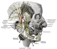
Photo from wikipedia
The purpose of this study is to evaluate the relationship between anatomic structures and mandibular posterior region using cone-beam computed tomography (CBCT) in terms of endodontic surgery. A total of… Click to show full abstract
The purpose of this study is to evaluate the relationship between anatomic structures and mandibular posterior region using cone-beam computed tomography (CBCT) in terms of endodontic surgery. A total of 150 CBCT images were used to investigate the proximity of the anatomical structures and the mandibular posterior teeth. The buccal and lingual bone thickness overlying each root, buccolingual, and mesiodistal dimension of the roots were measured at the level of 3 mm apical resection, and the mental foramen (MF) distance to the premolar teeth and the distance of the mandibular canal (MC) to all the posterior teeth were measured. The thinnest part of the buccal cortical bone was measured in the first premolar teeth (1.70 mm) and in the mesial root of the first molar (2.25 mm) while the thickest region was measured in the distal root of the second molar tooth (6.95 mm). The maximum amount of substance to be removed was measured at the distal root of the second molar tooth (11.26 mm), and at least the first premolar tooth (5.52 mm) was measured for buccal resection. The distal root of the second molar tooth was found to be the closest tooth root to the MC with a mean of 2.75 mm, and the closest distance was measured as 0 mm. It is important to evaluate the parameters such as mandibular buccal and lingual bone thickness, location of the MC and the MF, and root size for atraumatic endodontic surgical approach. Evaluation of these data before endodontic surgery provides guidance to the clinician in the planning of endodontic surgery. The mandibular posterior region, which is difficult to reach with traditional surgical approach, is now easily reached using an operation microscope. For this reason, endodontic surgical procedures have become popular in mandibular posterior teeth. Therefore, the relationship between the mandibular posterior teeth and anatomical structures that are important in the planning of surgical access line is examined in this study.
Journal Title: Clinical Oral Investigations
Year Published: 2019
Link to full text (if available)
Share on Social Media: Sign Up to like & get
recommendations!