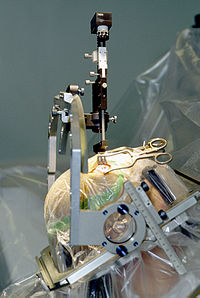
Photo from wikipedia
In recent years, with the development of new neurosurgical techniques and devices, increasingly advanced knowledge and techniques are needed to safely perform surgeries. Medical accidents caused by surgeons performing procedures… Click to show full abstract
In recent years, with the development of new neurosurgical techniques and devices, increasingly advanced knowledge and techniques are needed to safely perform surgeries. Medical accidents caused by surgeons performing procedures without sufficient experience have become a substantial challenge to society. Surgical training has always focused on learning in the operating room under the supervision of a senior surgeon. However, it is difficult to learn complicated anatomical structures and surgical approaches only by learning from the actual surgery. Therefore, there is an urgent need to build an educational programme for trainees with minimal risk to patients. We are very interested in the recent review article by Iwanaga et al. [5]. These authors analysed 37 cadaveric anatomical feasibility studies and investigated the required for the work to be clinically applied and published. The analysis showed that approximately 22% of the studies were clinically applied in the field of neurosurgery within 7 years after publication, and the median time until citation was 3.4 years [5]. In contrast, it has been reported that the time required for conventional clinical translational research to be applied clinically is 17 years [15]. Given that cadaveric anatomical studies are approximately 2.5 times faster than clinical translational research from the start of research to the treatment of patients, cadaveric research could be a potential strategy for advancing the field of neurosurgery. We applaud the work by Iwanaga et al. and thank these authors for providing valuable information. The importance and necessity of the cadaver laboratory in the education of neurosurgeons have been widely recognized internationally. Recently, even in developing countries, neurosurgical cadaver laboratories have begun to be established with limited medical resources [18, 19]. In addition to surgical training, new surgical approach development by cadaveric research has been carried out in cadaver laboratories [20, 21]. On the other hand, there is not a large number of cadavers available in each country [2, 3]. Recently developed three-dimensional (3D) print models may be promising solutions to the global shortage of cadavers [13]. In particular, disease-specific models such as those with cerebral aneurysms and brain tumours are becoming more accurate and are promising methods for preoperative simulation from a realistic perspective [1, 10, 11, 22]. However, due to the difficult construction of materials, some prototypes lacked brain tissue, and some used animal brain tissue [4, 23]. The 3D model has not been sufficiently verified for its usefulness in comparison with the educational tools such as two-dimensional models developed for residents. In addition, multifaceted evaluations such as anatomical accuracy, usefulness as a surgical training model, and cost for new 3D models should be continued, and comparison with CST should be performed. In Japan, the Japan Surgical Society (JSS) cooperated with the Japanese Association of Anatomists (JAA) and drafted the “Guidelines for Cadaver Dissection in Education and Research of Clinical Medicine” in 2012. With the establishment of a special committee by the JSS, all universities hosting cadaver surgical training (CST) are obligated to report the content of their CST implementation and their budgetary investment each year, considering the ethical importance of CST. Since this guideline was implemented, programmes for CST and organizations conducting cadaver * Yoshio Araki [email protected]
Journal Title: Neurosurgical Review
Year Published: 2022
Link to full text (if available)
Share on Social Media: Sign Up to like & get
recommendations!