Photo from wikipedia
Papaya (Carica papaya L.) is susceptible to viral diseases caused by Papaya mosaic virus (PapMV) and Papaya ringspot virus (PRSV), which limit fruit production and affect economic yield. The symptoms… Click to show full abstract
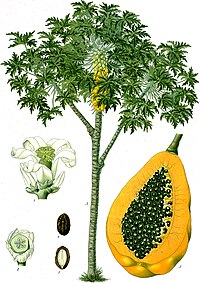
Papaya (Carica papaya L.) is susceptible to viral diseases caused by Papaya mosaic virus (PapMV) and Papaya ringspot virus (PRSV), which limit fruit production and affect economic yield. The symptoms produced by both the viruses are similar in early stages of infection and include vein and leaf chlorosis, which develop into mosaic at later stages of infection when leaf lamina can get reduced in size and distorted with a shoe-string aspect. Digital image analyses, such as fractal dimension (FD) and lacunarity (λ) were used here to examine papaya tissue after single and mixed infections of PRSV and PapMV. Morphological changes, such as hypoplasia, hyperplasia, and neoplasia are described in tissues with viral infections. Furthermore, we quantified these changes and suggest three ranges of tissue damage according to their λ values in rank 1 (0.01 to 0.39), rank 2 (0.4 to 0.79), and rank 3 (0.8 to 1). Our analyses suggest that in synergism and antagonism there is a competition of both viruses to occupy the same mesophyll cells, as indicated by their intermediate values of lacunarity.
Journal Title: Biologia Plantarum
Year Published: 2017
Link to full text (if available)
Share on Social Media: Sign Up to like & get
recommendations!