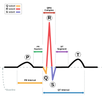
Photo from wikipedia
We report the case of a 26-year-old male with no structural cardiac abnormalities referred to our institution because of recurrent palpitations and a documented episode of a wide QRS tachycardia,… Click to show full abstract
We report the case of a 26-year-old male with no structural cardiac abnormalities referred to our institution because of recurrent palpitations and a documented episode of a wide QRS tachycardia, with right bundle branch blockmorphology, and left-axis deviation (Fig. 1a). Under isoproterenol infusion, a left posterior fascicular ventricular tachycardia (VT) with a cycle length of 300 ms was induced by programmed ventricular stimulation. Entrainment of the tachycardia was unsuccessfully attempted. The mechanism of fascicular VT is a localized reentry circuit involving ventricular myocardium immediately adjacent to the fascicle of the left bundle branch. In this case, the left ventricle was reached using the transseptal approach in combination with a long deflectable sheath. The left ventricular septum was electro-anatomically mapped during the tachycardia from the basal to the apical portion with a new 3D mapping system (Rhythmia, Boston Scientific, Boston, MA, USA), capable of rapidly acquiring detailed maps based on automatic annotation of thousands of points. The Rhythmia system acquired and automatically annotated 2500 electrograms over 95 s by the means of a bidirectional deflectable basket catheter with 64 closely spaced minielectrodes (IntellaMap OrionTM, Boston Scientific, Boston, MA, USA). The entire cycle length of the tachycardia was mapped by the system with automatic annotation and the earliest point of activation was identified at the mid posterior septum (Fig. 1 b, c). Two distinct signals preceding the ventricular electrogram were recorded by the OrionTM equatorial electrodes (Fig. 1d; A4–5/H4–5): the pre-Purkinje potential (P1) representing the verapamil sensitive zone activated anterogradely and a sharp signal which represents the Purkinje potential (P2), activated retrogradely. Thereafter, a manual modification of the window of interest was performed. The QRS complex was excluded, focusing exclusively on the diastolic part of the tachycardia (Fig. 1 e). This allowed us to identify the two limbs of the circuit on the activation map (Fig. 1 f; red indicates early activation; purple indicates late activation, the wave-front is * Giulio Conte [email protected]
Journal Title: Journal of Interventional Cardiac Electrophysiology
Year Published: 2017
Link to full text (if available)
Share on Social Media: Sign Up to like & get
recommendations!